Download
1 / 50
500 likes | 550 Views
Explore the complex genetics and wide range of diseases involving globin synthesis in thalassemias. Learn about major and minor variants, hemolysis, anemia types, and various hemolytic anemias like Megaloblastic Anemias. Delve into Iron Deficiency Anemia, Aplastic Anemias, and more.

E N D
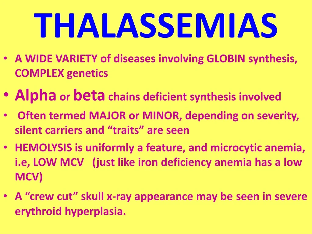
THALASSEMIAS • A WIDE VARIETY of diseases involving GLOBIN synthesis, COMPLEX genetics • Alpha or beta chains deficient synthesis involved • Often termed MAJOR or MINOR, depending on severity, silent carriers and “traits” are seen • HEMOLYSIS is uniformly a feature, and microcytic anemia, i.e, LOW MCV (just like iron deficiency anemia has a low MCV) • A “crew cut” skull x-ray appearance may beseen in severe erythroid hyperplasia.
Hemoglobin H Disease • Deletion of THREE alpha chain genes • HGB-H is primarilly Asian • HGB-H has a HIGH affinity for oxygen • HGB-H is unstable and therefore has classical hemolytic behavior
HYDROPS FETALIS • FOUR alpha chain genes are deleted, so this is the MOST SEVERE form of thalassemia • Many/most never make it to term • Children born will have a SEVERE hemolytic anemia as in the erythroblastosis fetalis of Rh disease: • Pallor (as in all anemias), jaundice, kernicterus • Edema (hence the name “hydrops”) • Massive hepatosplenomegaly (hemolysis)
Paroxysmal Nocturnal Hemoglobinuria (PNH) • ACQUIRED, NOT INHERITED like all the previous hemolytic anemias were • ACQUIRED mutations in phosphatidylinositol glycan A (PIGA) • Note: It is “P” and “N” only 25% of the time! GlycosylphosPhatidylInositol (lipid rafts)
Immunohemolytic Anemia • All of these have the presence of antibodies and/or compliment present on RBC surfaces • NOT all are AUTOimmune, some are caused by drugs • Antibodies can be • WARM (IgG) • COLDAGGLUTININ (IgM) • COLD HEMOLYSIN (paroxysmal) (IgG)
IMMUNOHEMOLYTIC ANEMIAS • WARM AGGLUTININS (IgG), will NOT agglutinate at room temp • Primary Idiopathic (most common) • Secondary (Tumors, especially leuk/lymph, drugs) • COLD AGGLUTININS: (IgM), WILL agglutinate at room temp • Mycoplasma pneumoniae, HIV, mononucleosis • COLD HEMOLYSINS: (IgG) Cold Paroxysmal Hemoglobinuria, hemo-LYSIS in body, ALSO often follows mycoplasma pneumoniae
COOMBSTEST • DIRECT: Patient’s CELLS are tested for surface Ab’s • INDIRECT: Patient’s SERUM is tested for Ab’s.
HEMOLYSIS/HEMOLYTIC ANEMIAS DUE TO RBC TRAUMA • Mechanical heart valves breaking RBC’s • MICROANGIOPATHIES: • TTP • Hemolytic Uremic Syndrome
NON-Hemolytic Anemias:i.e., DE-creased Production • “Megaloblastic” Anemias B12 Deficiency (Pernicious Anemia) Folate Deficiency • Iron Deficiency • Anemia of Chronic Disease • Aplastic Anemia • “Pure” Red Cell Aplasia • OTHER forms of Marrow Failure
MEGALOBLASTIC ANEMIAS • Differentiating megaloblasts (marrow) from macrocytes (peripheral smear, MCV>94) • Impaired DNA synthesis • For all practical purposes, also called the anemias of B12 and FOLATE deficiency • Often VERY hyperplastic/hypercellular marrow
Vit-B12 Physiology • Oral ingestion • Combines with INTRINSIC FACTOR in the gastric mucosa • Absorbed in the terminal ileum • DEFECTS at ANY of these sites can produce a MEGALOBLASTIC anemia
ALL megaloblastic anemias are also MACROCYTIC (MCV>94 or MCV~100), and that not only are the RBC’s BIG and hyperplastic/hypercellular, but so are the neutrophils, and neutrophilic precursors in the bone marrow too, and even more so, HYPERSEGMENTED!!!
PERNICIOUS ANEMIA • MEGALOBLASTIC anemia • LEUKOPENIA and HYPERSEGS • JAUNDICE • NEUROLOGIC posterolateral spinal tracts • ACHLORHYDRIA • Can’t absorb B12 • LOW serum B12 • Flunk Schilling test, i.e., can’t absorb B12, using a radioactive tracer
FOLATE DEFICIENCY MEGALOBLASTIC AMEMIAS • Decreased Intake: diet, infancy • Impaired Absorption: intestinal disease • DRUGS: anticonvulsants, BCPs, CHEMO • Increased Loss: Hemodialysis • Increased Requirement: Pregnancy, infancy • Impaired Usage
Fe Deficiency Anemia • Due to increased loss or decreased ingestion, almost always, nowadays, increased loss is the reason • Microcytic (low MCV), Hypochromic (low MCHC) • THE ONLY WAY WE CAN LOSE IRON IS BY LOSING BLOOD, because FE is recycled!
Fe Transferrin Ferritin (GREAT test) Hemosiderin
Clinical Fe-Defic-Anemia • Adult men: GI Blood Loss • PRE menopausal women: menorrhagia • POST menopausal women: GI Blood Loss
2 BEST lab tests: • Serum Ferritin • Prussian blue hemosiderin stain of marrow (also called an “iron” stain)
Anemia of Chronic Disease* • CHRONIC INFECTIONS • CHRONIC IMMUNE DISORDERS • NEOPLASMS • LIVER, KIDNEY failure * Please remember these patients may very very much look like iron deficiency anemia, BUT, they have ABUNDANT STAINABLE HEMOSIDERIN in the marrow!
APLASTIC ANEMIAS • ALMOST ALWAYS involve platelet and WBC suppression as well • Some are idiopathic, but MOST are related to drugs, radiation • FANCONI’s ANEMIA is the only one that is inherited, and NOT acquired • Act at STEM CELL level, except for “pure” red cell aplasia
APLASTIC ANEMIAS • CHLORAMPHENICOL • OTHER ANTIBIOTICS • CHEMO • INSECTICIDES • VIRUSES • EBV • HEPATITIS
MYELOPHTHISIC ANEMIAS • Are anemias caused by metastatic tumor cells replacing the bone marrow extensively
POLYCYTHEMIA • Relative (e.g., hemoconcentration) • Absolute • POLYCYTHEMIA VERA(Primary) (LOW EPO) • POLYCYTHEMIA (Secondary) (HIGH EPO) • HIGH ALTITUDE • EPO TUMORS • EPO “Doping”
P. VERA • A “myeloproliferative” disease • ALL cell lines are increased, not just RBCs
BLEEDING DISORDERS(aka, Hemorrhagic “DIATHESES”) • Blood vessel wall abnormalities √ • Reduced platelets √ • Decreased platelet function √ • Abnormal clotting factors √ • DIC (Disseminated INTRA-vascular Coagulation), also has ↓ plats.
VESSEL WALL ABNORMALITIES(angiopathic thrombocytopenias)(NON-thrombotic cytopenic purpuras) • Infections, especially, meningococcemia, and rickettsia • Drug reactions causing a leukocytoclastic vasculitis • Scurvy, Ehlers-Danlos, Cushing syndrome • Henoch-Schönlein purpura (mesangial IgA deposits too) • Hereditary hemorrhagic telangiectasia (Osler–Weber–Rendu syndrome, Autosomal Dominant) • Amyloid
THROMBOCYTOPENIAS • Like RBCs: • DE-creased production • IN-creased destruction • Sequestration (Hypersplenism) • Dilutional • Normal value 150K-300K
DE-CREASED PRODUCTION • APLASTIC ANEMIA • ACUTE LEUKEMIAS • ALCOHOL, THIAZIDES, CHEMO • MEASLES, HIV • MEGALOBLASTIC ANEMIAS • MYELODYSPLASTIC SYNDROMES (PRE-Leukemias)
IN-CREASED DESTRUCTION • AUTOIMMUNE (ITP) • POST-TRANSFUSION (NEONATAL) • QUINIDINE, HEPARIN, SULFA • MONO, HIV • DIC, “CONSUMPTIVE” • TTP/HUS • “MICROANGIOPATHIC”
THROMBOCYTOPENIAS • ITP (Idiopathic Thrombocytopenic Purpura) • Acute Immune • DRUG-induced • HIV associated • TTP, Hemolytic Uremic Syndrome
I.T.P. • ADULTS AND ELDERLY • ACUTE OR CHRONIC • AUTO-IMMUNE • ANTI-PLATELET ANTIBODIES PRESENT • INCREASED MARROW MEGAKARYOCYTES • Rx: STEROIDS
ACUTE ITP • CHILDREN • Follows a VIRAL illness (~ 2 weeks) • ALSO have anti-platelet antibodies • Platelets usually return to normal in a few months
DRUGS • Quinine • Quinidine • Sulfonamide antibiotics • HEPARIN
HIV • BOTH DE-creased production AND IN-creased destruction factors are present
Thrombotic Microangiopathies • BOTH are very SERIOUS CONDITIONS with a HIGH mortality: • TTP (THROMBOTIC THROMBOCYTOPENIC PURPURA) • H.U.S. (HEMOLYTIC UREMIC SYNDROME) • These can also be called “consumptive” coagulopathies, just like a DIC
“QUALITATIVE” platelet disorders • Mostly congenital (genetic): • Bernard-Soulier syndrome (Glycoprotein-1-b deficiency) • Glanzmann’s thrombasthenia (Glyc.-IIB/IIIA deficiency) • Storage pool disorders, i.e., platelets mis-function AFTER they degranulate • ACQUIRED: ASPIRIN, ASPIRIN, ASPIRIN
BLEEDING DISORDERS due toCLOTTING FACTOR DEFICIENCIES • NOT spontaneous, but following surgery or trauma • ALL factor deficiencies are possible • Factor VIII and IX both are the classic X-linked recessive hemophilias, A and B, respectively • ACQUIRED disorders often due to Vitamin-K deficiencies (II, VII, IX, X) • von Willebrand disease the most common, 1%
vonWillebrand Disease • 1% prevalence, most common bleeding disorder • Spontaneous and wound bleeding • Usually autosomal dominant • Gazillions of variants, genetics even more complex • Prolonged BLEEDING TIME, NL platelet count • vWF is von Willebrand Factor, which complexes with Factor VIII, it is the von Willebrand Factor which is defective in von Willebrand disease • Usually BOTH platelet and FactorVIII-vWF disorders are present
PTT PT/INR
HEMOPHILIA A • The “classic” HEMOPHILIA • Factor VIII decreased • Co-factor of Factor IX to activate Factor X • Sex-linked recessive • Hemorrhage usually NOT spontaneous • Wide variety of severities • Prolonged PTT (intrinsic) only • Rx: Recombinant Factor VIII
HEMOPHILIA B • The “Christmas” HEMOPHILIA • Factor IX decreased • Sex-linked recessive • Hemorrhage usually NOT spontaneous • Wide variety of severities • Prolonged PTT (intrinsic) only • Rx: Recombinant Factor IX
DIC, Disseminated INTRA-vascular, Coagulation • ENDOTHELIAL INJURY • WIDESPREAD FIBRIN DEPOSITION • HIGH MORTALITY • ALL MAJOR ORGANS COMMONLY INVOLVED
DIC, Disseminated INTRA-vascular, Coagulation • Extremely SERIOUS condition • NOT a disease in itself but secondary to many conditions • Obstetric: MAJOR OB complications, toxemia, sepsis, abruption • Infections: Gm-, meningococcemia, RMSF, fungi, Malaria • Many neoplasms, acute promyelocytic leukemia • Massive tissue injury: trauma, burns, surgery • “Consumptive”coagulopathy
Common Coagulation TESTS • PTT (intrinsic) • PT INR (extrinsic) • Platelet count, aggregation • Bleeding Time, so EASY to do • Fibrinogen • Factor Assays
RBC LAB http://www.chronolab.com/hematology/2_1.htm