Download
1 / 18
180 likes | 261 Views
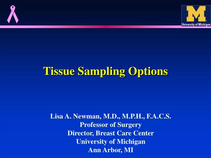
Tissue Sampling Options. Lisa A. Newman, M.D., M.P.H., F.A.C.S. Professor of Surgery Director, Breast Care Center University of Michigan Ann Arbor, MI. Breast Masses/Abnormalities. Palpable or Non-Palpable (screen-detected) Benign or Cancerous

E N D
Tissue Sampling Options Lisa A. Newman, M.D., M.P.H., F.A.C.S. Professor of Surgery Director, Breast Care Center University of Michigan Ann Arbor, MI
Breast Masses/Abnormalities • Palpable or Non-Palpable (screen-detected) • Benign or Cancerous • Benign: Cyst; Fibroadenoma; Fibrocystic Density • Breast imaging (mammo and sono) can identify the benign masses frequently, but not always! • Biopsy is ALWAYS necessary to document cancer, and is frequently necessary to confirm benign lesions • Whenever possible, needle biopsies are preferred as the first step in diagnosing a breast mass
Considerations when Choosing Biopsy Technique • Available resources • Surgical services • Technology; biopsy devices • Pathology interpretive services • Goal of biopsy material • Establish diagnosis • For cancers, must have adequate tissue for therapeutic marker informatioin • ER; PR; HER2/neu • Research/tissue banking?
Considerations when Choosing Biopsy Technique • Biopsy result MUST be concordant with clinical impression • If not, then additional sampling and/or additional pathoology review is necessary • Patient follow-up is essential!!!! • Multidisciplinary management of breast lesions involves pathology • Must plan biopsy program with path based on local resources and expertise
HISTOPATHOLOGIC WORK-UPOF PALPABLE MASSES • Open, excisional biopsy (gold standard) • Open, incisional biopsy • Percutaneous, free-hand needle biopsy • Fine-needle aspirate (FNA) • Core, Tru-Cut needle biopsy • Image-directed needle biopsy • Ultrasound-guided • Stereotactic/mammo-guided
HISTOPATHOLOGIC WORK-UPOF PALPABLE MASSES • Open, excisional biopsy (gold standard) vs. Open, incisional biopsy • Both require use of surgical services • Excisional implies complete removal of mass • Incisional useful with large, bulky lesions • Advantageous in pts receiving neoadjuvant chemotherapy • Either approach provides substantial amounts of tissue for diagnosis, molecular marker analyses
HISTOPATHOLOGIC WORK-UPOF PALPABLE MASSES • Percutaneous, free-hand needle biopsy • Fine-needle aspirate (FNA) • Core, Tru-Cut needle biopsy • FNA: readily-available small-guage needles can obtain material for cytologic analyses • No tissue wedge available and therefore cannot reliably distinguish invasive from in situ disease • Very useful for evaluation of bulky lymph nodes • Requires additional cytopathologic expertise for appropriate interpretation • Requires additional pathology expertise in order to obtain molecular marker information (ER/PR/HER2)
HISTOPATHOLOGIC WORK-UPOF PALPABLE MASSES • Percutaneous, free-hand needle biopsy • Fine-needle aspirate (FNA) • Core, Tru-Cut needle biopsy • FNA: readily-available small-guage needles can obtain material for cytologic analyses (22-26 G) • Easily-tolerated by patient • No tissue wedge available and therefore cannot reliably distinguish invasive from in situ disease • Requires additional cytopathologic expertise for appropriate interpretation • Preferable to have special storage solution for specimen transport and processing • Requires additional pathology expertise in order to obtain molecular marker information (ER/PR/HER2)
HISTOPATHOLOGIC WORK-UPOF PALPABLE MASSES • Percutaneous, free-hand needle biopsy • Fine-needle aspirate (FNA) • Core, Tru-Cut needle biopsy • Core needle biopsy: commercially-available, spring-loaded needle biopsy devices (14-18 Guage; 10 or 20 mm length cores of tissue extracted) • Requires local anesthetic; multiple passes • Specimen cores placed in formalin solution for transport and processing • Adequate tissue for full histopath architecture evaluation and molecular marker studies • Adequate tissue for tumor banking
KATH-UM Breast Cancer Collaboration Core Needle Biopsies at KATH • Prior to summer 2008: majority of breast tumors at KATH evaluated by either surgical biopsy or FNA biopsy • KATH staff concerned about inefficient use of surgical resources and tissue adequacy with FNA biopsies • July 2008: Training course implemented
KATH-UM Breast Cancer Collaboration Core Needle Biopsies at KATH • 2006-2008: UM and KATH teams work in clinic together to evaluate breast masses • July 2008: Training course implemented • Since training course implemented, KATH pathology available on 82 core biopsy specimens • UM path review on malignant 46 cores, all confirming cancer • UM path review on 10 benign cores, all concordant by UM review (mostly fibroadenoma diagnosis)
HISTOPATHOLOGIC WORK-UPOF PALPABLE MASSES • Open, excisional biopsy (gold standard) • Open, incisional biopsy • Percutaneous, free-hand needle biopsy • Fine-needle aspirate (FNA) • Core, Tru-Cut needle biopsy • Image-directed needle biopsy • Ultrasound-guided • Stereotactic/mammo-guided • Can minimize risk of sampling error/false-negative result • Requires mammography and ultrasound equipment; radiology expertise
HISTOPATHOLOGIC WORK-UPOF PALPABLE MASSES • Open, excisional biopsy (gold standard) • Open, incisional biopsy • Percutaneous, free-hand needle biopsy • Fine-needle aspirate (FNA) • Core, Tru-Cut needle biopsy • Image-directed needle biopsy • Ultrasound-guided • Stereotactic/mammo-guided • Punch Biopsy Device: Circular scalpel • Classically utilized by dermatologists • Easy-to-perform, office-based procedure under local anesthesia • Useful for breast lesions involving skin • 3; 4; and 6 mm punch biopsy devices available • Will usually require suture to close remaining skin defect
Wire localization surgical biopsy for mammographically-detected breast lesion
Summary • Multiple biopsy options are available • Surgical resection is gold standard for complete histopathologic evaluation • Percutaneous needle biopsies are efficient and spare surgical rescources • Punch biopsies useful for ulcerated lesions involving skin • Multidisciplinary management of breast lesions involves pathology • Must plan biopsy program with path based on local resources and expertise • CONCORDANCE is critical!!!! • Hand-in-hand with patient follow-up