Download
1 / 98
980 likes | 1.12k Views
C hair of M icrobiology, V irology, and I mmunology. PATHOGENIC AND CONDITIONALLY-PATHOGENIC ENTERIC GRAM-NEGATIVE BACTERIA. ESCHERICHIA. SALMONELLA. Lecturer Prof. S.I. Klymnyuk.

E N D
Chair of Microbiology, Virology, and Immunology PATHOGENIC AND CONDITIONALLY-PATHOGENIC ENTERIC GRAM-NEGATIVE BACTERIA. ESCHERICHIA. SALMONELLA. Lecturer Prof. S.I. Klymnyuk
The Enterobacteriaceae contain gram negative rodswhich, if motile, are peritrichously flagellated. Because members of this family are morphologically and metabolically similar, much effort has been expended to develop techniques for their rapid identification. In general, biochemical properties are used to define a genus, and further subdivision frequently is based on sugar fermentation and antigenic differences. Yet, many paradoxes exist, for example, more than 2200 species of Salmonella have been named, whereas the equally complex species Escherichia coli is divided into more than 1000 serotypes.Over the years, many taxonomists with different ideas have been involved in the classification of these bacteria, and disagreement still exists concerning family and generic names.
Biochemical Properties Used for Classification Early taxonomic schemes relied heavily on the organism's ability to ferment lactose, and numerous differential and selective media have been devised to allow one to recognize a lactose fermenting colony on a solid medium. The effectiveness of such differential media is based on the fact that organisms fermenting the lactose form acid, whereas nonlactose fermenters use the peptones present and do not form acids m these media. The incorporation of an acid base indicator into the agar medium thus causes a color change around a lactose fermenting colony. Thus has been a valuable technique for selecting the major non lactose fermenting pathogens that cause salmonellosis or shigellosis, under special conditions, however, many lactose fermenters also cause a variety of infectious diseases.
Furthermore, many enterics ferment lactose only slowly, requiring several days before sufficient acid is formed to change the indicator. They all synthesize beta galactosidase, (the enzyme that splits lactose into glucose and galactose) but lack the specific permease necessary for the transport of lactose into the cell One can easily determine whether an organism is a slow lactose or nonlactose fermenter by mixing a loopful of bacteria with orthonitrophenol beta galactoside (ONPG) dissolved in a detergent. The linkage of the galactose in ONPG is the same as its linkage m lactose, inasmuch as the ONPG can enter the cell in the absence of a permease, an organism possessing beta galactosidase will hydrolyze ONPG to yield galactose and the bright yellow compound, orthonitrophenol. Thus, only the absence of a specific lactose permease differentiates the slow lactose fermenters from the normal lactose fermenters.
In addition, a number of selective media have been devised that contain bile salts, dyes such as brilliant green and methylene blue, and chemicals such as selenite and bismuth. The incorporation of such compounds into the growth of medium has allowed for the selective growth of the enterics while inhibiting the growth of gram positive organisms. Some other biochemical properties used to classify members of the Enterobacteriaceae include the ability to form H2S; decarboxylate the ammo acids lysine, ornithine,or phenylalanine, hydrolyze urea intoCO2 and NH3, form indole from tryptophan; grow with citrate as a sole source of carbon; liquefy gelatin; and ferment a large variety of sugars. Serologic Properties Used for Classification No other group of organisms has been so extensively classified on the basis of cell surface antigens as the Enterobacteriaceae. These antigens can be divided into three types, designated O, K, and H antigens.
O ANTIGENS. All gram-negative bacteria possess a lipopolysaccharide (LPS) as a component of their outer membrane. This toxic LPS (also called endotoxin) is composed of three regions, lipid A, core, and are peating sequence of carbohydrates called the O antigen. Based on different sugars, alpha- or beta-glycosidic linkages, and the presence or absence of substituted acetyl groups, Escherichia coil can be shown to possess at least 173 different O antigens, and 64 have been described in the genus Salmonella. Sometimes, after continuous laboratory growth, strains will, through mutation, lose the ability to synthesize or attach this oligosaccharide O antigen to the core region of the LPS. This loss results in a change from a smooth colony to a rough colony type, and it is referred to as an S to R transformation Interestingly, the R mutants have lost the ability to produce disease.
K ANTIGENS. K antigens exist as capsule or envelope polysaccharides and cover the O antigens when present, inhibiting agglutination by specific O antiserum.Most K antigens can be removed by boiling the organisms in water.
H ANTIGENS. Only organisms that are motile possess H antigens because these determinants are in the proteins that makeup the flagella. However, to complicate matters, members of the genus Salmonella alternate back and forth to form different H antigens. The more specific antigens are called phase 1 antigens and are designated by lower-case letters (a, b, c, and so on), whereas the less-specific phase 2 H antigens are given numbers. The mechanism of this phase variation reveals an interesting way in which a cell canregulate the expression of its genes. In short, Salmonella possesses two genes. H1 encoding for phase 1 flagellar antigens, and H2 encoding for phase 2 flagellar antigens.The transcription of H2 results in the co-ordinate expression of gene rhl, which codes for a repressor that preventsthe expression of H 1.
About every 103 to 10s generations, a 900-base-pair region, containing the promoter for the H2 gene, undergoes a site-specific inversion, stopping the transcription of both H2 and rhl. In the absence of the rhl gene product, the H1 gene is then transcribed until the 900-base pair region in the H2 promoter is again inverted, resulting in the expression ofH2 and rhl. After obtaining the serologic data, an antigenic formula can be written, such as E.coli O111:K-58:H6, meaning this E. coli possesses O antigen 111, K antigen 58, and H antigen 6. The formula Salmonella to go 4,12:1,w:1,6 indicates this serotype of Salmonella possesses O antigens 4 and 12, phase 1 H antigens 1 and w, and phase 2 H antigens 1 and 6.
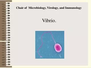
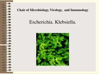
Escherichia coli. The organism was isolated from faeces in 1885 by T. Escherich. E. coli is a common inhabitant of the large intestine of humans and mammals. It is also found in the guts of birds, reptiles, amphibians, and insects. The bacteria are excreted in great numbers with the faeces and are always present in the external environment (soil, water, foodstuffs, and other objects). Morphology. E coli are straight rods measuring 0.4-0.7 in breadth and 1-3 in length. They occur as individual organisms or in pairs and are marked by polymorphism. There are motile and non-motile types. The G+C content in DNA is 50-51 per cent. The cell surface has pili on which certain phages are adsorbed. The microcapsule is not always clearly defined.
Cultivation. E. coli is a facultative anaerobe. The optimum temperature for growth is 30-37 °C and the optimum pH value of medium up 7.2-7.5. The organism also grows readily on ordinary media at room temperature and at 10 and 45 °C, growth becomes visible in the first two days. E. coli from cold-blooded animals grows at 22-37° C but not at 42-43° C. On meat-peptone agar E. coli produces slightly convex semitransparent, greyish colonies, and in meat broth it forms diffuse turbidity and a precipitate. The organism produces colonies which are red on Ploskirev's medium, red with a metallic hue on Endo's medium, and dark-blue on Levin's medium.
Fermentative properties. E. coli does not liquefy gelatin. It produces indole and hydrogen sulphide, and reduces nitrates to nitrites; ferments glucose, levulose, lactose, maltose, mannitol, arabinose, galactose, xylose, rhamnose, and occasionally saccharose, raffinose, dulcitol, salycin, and glycerin, with acid and gas formation. It also coagulates milk. There are varieties of the bacteria which ferment saccharose, do not produce indole, have no flagella, and do not ferment lactose.
Toxin production. Certain strains of E. coli are conditionally pathogenic They contain a gluco-lipo-protein complex with which their toxic, antigenic, and immunogenic properties are associated. Some strains possess haemolytic properties (O124 and others) determined by plasmids. Pathogenic cultures possess endotoxins and thermolabile neurotropic exotoxins. The latter accumulate in broth cultures on the second-fourth day of cultivation, while the endotoxins appear only after the twentieth day. Haemotoxins and pyrogenic substances, proteinases, deoxyribonucleases, urease, phosphatase, hyaluronidase, amino acid decarboxylases have been obtained from pathogenic strains.
Antigenic structure. The antigenic structure of E. coli is characterized by variability and marked individuality. Along with the H- and O-antigens, the presence of other antigens has been shown m some strains, i.e. the surface somatic (membranous, capsular) K-antigens which contain the thermolabile L- and B-antigens and the thermostable A- and M-antigens. Each antigen group in its turn is composed of a number of antigens designated by Arabic numbers, e.g. the O-group has 173 antigens, the K-subgroup 90, the H-subgroup 50, etc. On the basis of antigenic structure an antigenic formula is derived which fully reflects the antigenic properties of the strain For example, one of the most widely spread serotypes is designated 0111 : K58 : H2. Under the effect of transformation, lysogenic conversion, transduction, and conjugation E. coli may change its antigenic properties. Numerous varieties of the organism are produced on cultivation under artificial conditions. Such varieties are not only of theoretical interest, but also of great practical importance in laboratory diagnosis of enteric infections.
Classification. Genus Escherichia includes one E. coli species consisting of several biotypes and serotypes. They are differentiated according to cultural, biochemical, and serological properties. The genus Escherichia includes E. coli, E. freundi, E. vulneris, and others. E. coli comprises several varieties which are differentiated by their cultural and biochemical properties. F. Kauffmann has detected 25 O-groups responsible for various diseases in humans. About 50 phage variants have been revealed among E. coli organisms. They are used in laboratory diagnosis as confirmatory characteristics of the isolated serotypes.
Resistance. E. coli survives in the external environment for months. It is more resistant to physical and chemical factors of the external environment than the typhoid and dysentery bacteria. E. coli is killed comparatively rapidly by all methods and preparations used for disinfection. At 55° C the organism perishes in 1 hour, and at 60° C in 15 minutes. E. coli is sensitive to brilliant green. E. coli is used as a test microbe in the assay of disinfectants and methods of disinfection and also in titration of certain antibiotics. Pathogenicity for animals. The pathogenic serovars of E. coli cause severe infections in calf sucklings giving rise to an extremely high mortality. A parenteral injection of the culture into rabbits, guinea pigs, and white mice results in a fatal toxico-septical condition.
Pathogenesis and diseases in man. Definite E. coli serogroups are capable of causing various acute intestinal diseases in humans: (1) the causative agents of colienteritis in children are O-groups-25, -26, -44, -55, -86, -91, -111, -114, -119, -125, -126, -127, -128, -141, -146, and others (they cause diseases in infants of the first months of life and in older infants); (2) the causative agents of dysentery-like diseases are E. coli of the O-groups-23, -32, -115, -124, -136, -143, -144, -151, and others; (3) the causative agents of cholera-like diarrhoea are the O-groups-6, -15, -78, -148, and others, they produce thermolabile and thermoresistant enterotoxins. Colienteritis begins acutely with high temperature (38-39 °C), and frequently with severe meteorism, vomiting, diarrhoea, and general toxicosis. The disease usually occurs in infants of the first year of life.
The infection is acquired from sick children or carriers. Pathogenic E. coli serovars are found on various objects. It is assumed that colienteritis is transmitted not only by the normal route for enteric infections but also through the respiratory tract by the droplets and dust. The pathogenesis of colienteritis depends entirely on the organism's condition. In prematurely born infants and in infants during the first months of life the bactericidal activity of blood is considerably lower in respect to the pathogenic E. coli serovars in comparison to the nonpathogenic types. The reactivity of the child's body at the time of infection plays an important role in the mechanism of resistance to the pathogenic strains.
The pathological process develops mainly in the small intestine. Most probably, the mucous membrane of the small intestine in particular is exposed to the action of thermolabile toxic substances. Serovars O-124, O-151 and others cause diseases which are similar to dysentery. E. coli may cause colibacillosis in adults (peritonitis, meningitis, enteritis, toxinfections, cystitis, pyelitis, pyelonephntis, angiocholitis, salpingooophontis, appendicitis, otitis, puerperal sepsis, etc.). Over-strain, exhaustion, and conditions following infectious diseases facilitate the onset of various E. coli infections. In a number of cases the organism is responsible for food poisoning.
Immunity. In individuals who had suffered from diseases caused by pathogenic E. coli serovars, cross immunity is not produced owing to which re-infection may occur. Over 85 per cent of E. coli strains contain inhibiting substances, colicins, marked by antagonistic properties in relation to pathogenic microbes of the enteric group, they are used as therapeutic and preventive agents, e.g. colibacterin (E. coli M 17, etc.). Besides this, E colt as well as other common inhabitants of the intestine are capable of synthesizing various vitamins (K2, E, and group B) which are indispensable to the human organism. The ability of various E. coli serovars to suppress the growth of Mycobacterium tuberculosis has also been observed. The suppression of E. coli and other members of the biocoenosis may result in a chronic disease known as dysbacteriosis.
Laboratory diagnosis. The patients' faeces, throat and nasal discharges, material obtained at autopsy (blood, bile, liver, spleen, lungs, contents of the small and large intestine, pus), water, foodstuffs, and samples of washings from objects and hands of staff of maternity hospitals, hospitals, and dairy kitchens are all used for laboratory examination during colienteritis. If possible, faecal material should be seeded immediately after it has been collected. The throat and nasal discharges are collected with a sterile swab. Specimens of organs obtained at autopsy are placed in separate sterile jars.
The tested material is inoculated onto solid nutrient media (Endo's, Levin's) and, simultaneously, onto Ploskirev's media and bismuth-sulphite agar for isolation of bacteria of the typhoid-paratyphoid and dysentery group. BIood is first inoculated into broth and then subcultured on solid media when development of a septic process is suspected. Pus is collected for examination in suppurative lesions. It is placed into a dry sterile vessel and then inoculated onto the differential media of Endo or Levin. The pure culture isolate is identified by its morphological, cultural, biochemical, serological, and biological properties.
The corresponding O-group to which an enteropathogenic-serovars belong is determined by means of the agglutination reaction after the K-antigen of the culture that is being studied has been destroyed by boiling. Besides, the immunofluorescence method employing type specific labelled sera is also used. It yields a preliminary answer in one to two hours. In serological diagnosis of colienteritis beginning with the third to fifth day of the disease the indirect haemagglutination reaction is used which excels the agglutination reaction in sensitivity. It is positive when the antibody titre grows in the course of the diseased.
Treatment. Patients with colienteritis are prescribed antibiotics (tetracycline with vitamins C, B1 and B2) and biopreparations (coli autovaccine, coli bacteriophage, colicin, bacterin, lactobacterin, bificol, bifidumbacterin). Physiological solutions with glucose are injected for controlling toxicosis. Prophylaxis. To prevent diseases caused by pathogenic serovars of E. coli, special attention is given to early identification of individuals suffering from colienteritis, and also to their hospitalization and effective treatment. Regular examination of personnel is necessary in children's institutions as well as of mothers whose children are suffering from dyspepsia. Considerable importance is assigned to observation of sanitary regulations in children's institutions, infant-feeding centres, maternity hospitals, and children's nurseries. Protection of water and foodstuff's from contamination with faeces, the control of flies, and gradual improvement of standards of hygiene of the population are also particularly important.
Sanitary significance of E. coli. This organism is widely spread in nature. It occurs in soil, water, foodstuff's, and on various objects. For this reason E. coli serves as an indicator of faecal contamination of the external environment. Detection of E. coli is of great importance in estimating the sanitary index of faecal contamination of water, foodstuff's, soil, beverages, objects, and hand-washings. The degree of contamination of water, soil and foodstuff's is determined by the coli titre or coli index (these terms have been discussed in the chapter concerning the spread of microbes in nature). Faecal contamination of articles of use is estimated by qualitative determination of the presence of E. coli.
Additional materials Pathogenicity of Escherichia coil. Although E. coli is part of the normal flora of the intestinal tract, it is also the most common gram-negative pathogen responsible for nosocomially acquired septic shock, meningitis in neonates, cystitis and pyelonephritis in women, and for several distinct forms of diarrheal disease and dysentery affecting populations throughout the world. Strains of E coli capable of causing such diseases possess one or more virulence factors that are not found in E. coli strains comprising the normal flora. Such virulence factors can be characterized as follows, the capacity to adhere to specific mammalian cells; the ability to invade and grow intracellularly in intestinal epithelial cells; the secretion of one or more enterotoxins that cause fluid loss, resulting diarrhea; the formation of a cytotoxin that blocks protein synthesis, causing a hemorrhagic colitis; and the possession of an antiphagocytic capsule that is responsible, at least in part, for the bacteremia and meningitis caused by E. coli. In addition, the ability to obtain iron from transferrin or lactoferrin by the synthesis of iron-binding siderophores markedly enhances the virulence of such strains through their ability to grow in host tissues.
Diarrheal Diseases. It is estimated that during the American Revolutionary War there were more deaths from diarrhea than from English bullets, and during the American War between the states, over 25% of all deaths were because of diarrhea and dysentery. Diarrhea kills more people worldwide than AIDS and cancer, with about five million diarrheal deaths occurring annually primarily because of dehydration. Most of these occur in neonates and young children, anda large number are caused by pathogenic E. coli. The disease in adults, known by many names such as traveller’s diarrhea or Montezuma's revenge, may vary from a milddisease with several days of loose stools to a severe and fatal cholera-like disease. Such life-threatening E. coli infections occur throughout the world but are most common in developing nations. The virulence factors responsible for diarrheal disease are frequently encoded in plasmids, which may be spread from one strain to another either through transduction: or by recombination. As a result, various combinations of virulence factors have occurred, which has been used to place the diarrhea-producing strains of E. coli into various groups based on the mechanism of disease production
Enterotoxigenic Escherichia coli. Enterotoxin-producing E coli, called enterotoxigenic E.coli (ETEC), produce one or both of two different toxins – a heat labile toxin called LT and a heat-stable toxin called ST. The genetic ability to produce both LT and ST is controlled by DNA residing in transmissible plasmids called ent plasmids. Both genes have been cloned, and the ST gene has been shown to possess the characteristics of a transposon.
HEAT-LABILE TOXIN. The heat-labile toxin LT, which is destroyed by heating at 65 °C for 30 minutes, has been extensively purified, and its mode of action is identical to that described for cholera toxin (CT). LT has a molecular weight of about 86,000 daltons and is composed of two subunits, A and B Subunit A consists of one molecule of Ai (24,000 daltons) and one molecule of A2 (5000daltons) linked by a disulfide bridge. Each A unit is joined noncovalently to five B subunits. Like CT, LT causes diarrhea by stimulating the activity of a membrane-bound adenylate cyclase.This results in the conversion of ATP to cyclic AMP (cAMP): ATP cAMP + PPi
Minute amounts of cAMIP induce the active secretion of Cl– and inhibit the absorption of NaCI, creating an electrolyte imbalance across the intestinal mucosa, resulting in the loss of copious quantities of fluid and electrolytes from the intestine. The mechanism by which LT stimulates the activity of the adenylate cyclase is as follows: (1) The B subunit of the toxin binds to a specific cell receptor,GM1 ganglioside, (2) the A1 subunit is released from the toxin and enters the cell; and (3) the A1 subunit cleaves nicotinamide-adenic dinucleonde (NAD) into nicotinamide and ADP-ribose and, together with a cellular ADP-ribosylating factor, transfers the ADP-ribose to aGTP-binding protein. The ADP-ribosylation of the GTP-binding protein inhibits a GTPase activity of the binding protein, leading to increased stability of the catalytic cornplex responsible for adenylate cyclase activity. This results in an amplified activity of the cyclase and a corresponding increase in the amount of cAMP produced.
Two antigenically distinct heat labile toxins are produced by various strains of E. coli. LT-I is structurally and antigenically related to CT to an extent that anti-CT will neutralize LT I LT-II has, on rare occasions, been isolated from the faeces of humans with diarrhea, but it is most frequently isolated from feces of water buftalos and cows LT-II is biologically similar to LT-I, but it is not neutralized by either anti-LT-I or anti-CT. LT will bind to many types of mammalian cells, and its ability to stimulate adenylate cyclase can be assayed in cell cultures. A report has also shown that CT stimulated an increase in prostaglandin E (PGE), and that PGE1 and PGE2 caused a marked fluid accumulation in the ligated lumen of rabbit intestinal segments. The mechanism whereby CT induces PGE release is unknown.
HEAT-STABLE TOXIN The heat-stable toxin STa consists of a family of small, heterogeneous polypeptides of 1500 to 2000 daltons that are not destroyed by heating at 100 °Cfor 30 minutes. STa has no effect on the concentration of cAMP, but it does cause a marked increase m thecellular levels of cyclic GMP (cGMP).cGMP causes an inhibition of the cotransport of NaCI across the intestinal wall, suggesting that the action of STa may be primarily antiabsorptive compared with that of LT, which is both antiabsorptive and secretory.
STa stimulates guanylate cyclase only in intestinal cells, indicating that such cells possess a unique receptor for STa. The cell receptor for STa is known to be either tightly coupled to, or a part of, a particulate form of guanylate cyclase located in the brush border membranes of intestinal mucosal cells. Also, intimately associated with this complex is a cGMP-dependent protein kinase that phosphorylates a 25,000 dalton protein in the brush border. It has been proposed that this phosphorylated protein might be the actual mediator for the toxin-induced ion transport alterations that lead to fluid loss. The usual assay for STa is to inject the toxin intragastrically into a 1 – to 4-day old suckling mouse and measure intestinal fluid accumulation (as a ratio of intestinal/remaining body weight) after 4 hours. STa may also be assayed directly by measuring its effect on the increase in guanylate cyclasein homogenized intestinal epithelial cells.
A second heat stable toxin that is produced by somestrains of E. coli has been termed STb. This toxin is inactivein suckling mice but will produce diarrhea in weaned piglets. STb producers have not been isolated from humans. It does not seem to increase the level of adenylate or guanylate cyclase in intestinal mucosal cells, but maystimulate the synthesis of prostaglandin E2. The end resuit is to enhance net bicarbonate ion secretion.
COLONIZATION FACTORS. Animals also are subject to infections by their own strains of ETEC, and such infections in newborn animals may result in death from the loss of fluids and electrolytes. Extensive studies of strains infecting newborn calves and piglets (as well as humans) have revealed that, in addition to producing an enterotoxin, such strains possess one of several fimbriate surface structures that specifically adhere to the epithelial cells lining the small intestine. These antigens (K-88 for swine strains, and K-99 for cattle) usually are fimbriate structures that cause the toxin-producing organisms to adhere to and colonize the small intestine. The need for this colonizing ability is supported by the fact that antibodies directed against the colonizing fimbriae are protective.
Analogous human ETEC strains also possess fimbriate structures that have been designated as colonizationfactors (CFA). At least five such serologically different factors, CFA/I, CFA/II, CFA/III, E8775, and CFA/V, have been described. Interestingly, these colonization factors also are plasmid mediated, and single plasmids have been described that carry genes for both CFA/I and STa. Interestingly, during the Gulf War in 1990, there were about 100 cases of diarrhea per week per 1000 personnel. Of these, 55% resulted from ETEC.
Enterohemorrhagic Escherichia coli. The enterohemorrhagic E. coli (EHEC) were first described in 1982 when they were shown to be the etiologic agent of hemorrhagic colitis, a disease characterized bysevere abdominal cramps and a copious, bloody diarrhea. These organisms are also known to cause a condition termed hemolytic-uremic syndrome (HUS), which is manifested by a hemolytic anemia, thrombocytopenia (decrease in the number of blood platelets), and acute renal failure. HUS occurs most frequently in children.
Although most initially recognized EHEC belong to serotype O157:H7, other EHEC serotypes such as O26, O111, O128, and O143 have been recognized. These organisms are not invasive, but they do possess a 60-megadalton plasmid that encodes for a fimbrial antigen that adheres to intestinal epithelium. In addition, the EHEC are lysogenic for one or more bacteriophages that encode for the production of one or both of two antigenically distinct toxins. These toxins are biologically identical and antigenically similar to the toxins formed by Shigella dysenteriae (Shiga's bacillus), and are designated as Shiga-like toxin I (SLT-I) and Shiga-like toxin II (SLT II). Because the Shiga-like toxins initially were characterized by their ability to kill Vero cells, a cell line developed from African green monkey kidney cells, they also arecalled Verotoxin I and Verotoxin II.