Download
1 / 28
280 likes | 566 Views
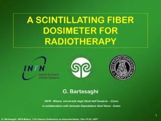
A SCINTILLATING FIBER DOSIMETER FOR RADIOTHERAPY. G. Bartesaghi. INFN - Milano, Università degli Studi dell’Insubria - Como in collaboration with Azienda Ospedaliera Sant’Anna - Como. Radiotherapy in the cancer care Why a multichannel scintillator detector?

E N D
A SCINTILLATING FIBER DOSIMETER FOR RADIOTHERAPY G. Bartesaghi INFN - Milano, Università degli Studi dell’Insubria - Como in collaboration with Azienda Ospedaliera Sant’Anna - Como
Radiotherapy in the cancer care Why a multichannel scintillator detector? Single scintillating fiber prototype Multifiber monodimensional detector 2D prototype Conclusions and outlooks Outline This work is part of the project PhoNeS (Photon Neutron Source) by INFN supported by MIUR PRI 05
Radiotherapy in the cancer care In the last decades, a constant progress has been made in the fight against cancer Data from EUROCARE 3 (European cancer registries study on cancer patients’ survival and care) Three factors have a crucial role: - primary prevention • earlier diagnosis • better treatment
General treatment methods } - chemotherapy - immunotherapy systemic treatments } - radiotherapy - surgical removal loco-regional treatments Radiotherapy exploits the effect of ionizing radiation on human tissues to eliminate tumour cells or to control their development. The radiation beam typically consists in electrons or photons (energy up to 20 MeV) emitted by a linear accelerator. Radiotherapy is one of the most popular method in cancer treatment. Every year, more than 5 millions patients worldwide receive almost a radiotherapic treatment, exploiting more than 7500 linacs.
Quality assurance in Radiotherapy For the future, the goal for radiotherapy is the same of the past delivering a high dose in the target volume preserving the surrounding volume All the procedures that ensure consistency of medical prescription and safe fulfilment of that prescription are part of the quality assurance. The goal of this work is to develop of a dosimeter that, performing real time 2D measurement in the radiation beam, may simplify and accelerate the quality assurance controls.
Why a scintillator dosimeter? Most popular dosimeters: Ionization chambers and diodes real time, accurate single channel not tissue equivalent Radiographic films excellent 2D resolution long processing time not tissue equivalent TLD small size time consuming requiring great care
Why a scintillator dosimeter? A plastic multifiber scintillator dosimeter assures important advantages: - real time measurement of 2D dose distribution - high tissue equivalence - a large dynamic range (100 keV – more than 20 MeV) - excellent radiation hardness - no pressure and temperature dependence - no high voltage near the probe
Single channel dosimeter Assembled in order to test the method feasibility scintillating probe (diameter: 1mm, length: 10mm) white fiber BCF-98 (diameter: 1mm, length: 300mm) PMT white fiber BCF-98 (diameter: 1mm, length: 300mm) PMT High energy electrons crossing a transparent medium produce Cherenkov light, which is not proportional to absorbed dose. It is therefore necessary a twin detector, without the scintillating probe, in order to evaluate the Cherenkov contribution.
Linear Accelerator Clinac Varian 2100 Radiotherapy Unit Ospedale S. Anna Como (Italy) Electron beam: Energy: 6, 9, 12, 16, 20 MeV Dose rate: from 100 to 600 MU/min Field: up to 25x25 cm2 Photon beam: Energy: 6, 18 MV Dose rate: from 100 to 600 MU/min Field: up to 40x40 cm2 1 Monitor Unit = 1 Gy at the build up (maximum dose region) Particles are emitted in bunches (100-300 Hz, depending on the dose rate)
Read out electronics The read out electronics is based on a charge integrating ADC (12 channels), with the trigger taken from the linac itself 3ms 7ms
Measurement set up Linac In order to test the scintillating dosimeter, a Depth Dose Scan is performed, the detector is inserted in a PMMA phantom Read out electronics 1 m Detector in PMMA phantom Treatment couch
Depth Dose Curves in Electron Beams Field size: 10x10cm2 Our measurements are compared with data taken by a diode Scanditronix DEB101 (1.25 mm3)
Depth Dose Curves in Electron Beams Field size: 10x10cm2
Depth Dose Curve in Photon Beams Field size: 10x10cm2
Crossline scan In a 6 MV photon beam, the detector is moved across the 10x10cm2 field.
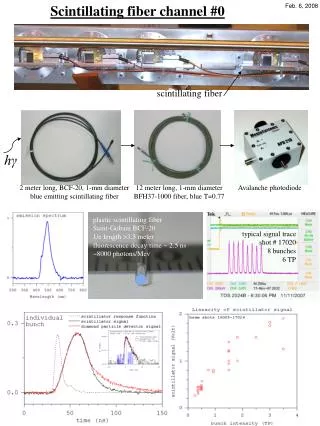
The single channel detector demonstrates the feasibility of a scintillating fiber dosimeter easy to handle and accurate. Next step is to develop a … Multifiber 1D prototype PMT 8 scintillating fibers (Bicron BCF-10, 1 cm long) inserted in a PMMA phantom are readout by a multichannel PMT. Each fiber is coupled with a twin white one for Cherenkov subtraction.
Intensity response The detector was positioned at different distances from the source in order to test the intensity response Data fitted with y = A/x2 + B
Reproducibility test 5 Gy are given 20 times Distribution of the charge read by the 5 scintillating fibers (each channel normalized to 100) The distribution RMS is about 0.5%
Depth Dose Curve in Photon Beams Field size: 10x10cm2
Depth Dose Curve in Electron Beams Field size: 10x10cm2
Crossline scan In a 6 MV photon beam, at 1.5 cm depth, the detector is moved across the 10x10cm2 field.
Gain equalization In order to evaluate the gain factor of the different channels the detector is positioned vertically and irradiated in a 6MV photon beam twice: from the top and from the bottom side The photon exponential attenuation is exploited to get the gain factors
Gain equalization … after the equalization Before and …
Multifiber 2D prototype 1 cm scintillating fiber + 1 cm white fiber in every pit 8 scintillating fibers are inserted vertically in a PMMA phantom (20x20x4 cm3) 16 white fibers (35 cm long) exiting from the bottom side of the phantom carry the light to the PMT
Depth Dose Scan Field size: 10x10cm2
Conclusions … - a scintillating fiber dosimeter can perform accurate real time measurement - good spatial resolution - good response at different radiation intensities - excellent reproducibility … and Outlooks - development of the 2D dosimeter increasing the number of channels (up to 256 or more) - some equalization methods are under evaluation - development of an integrated electronics (connection to PC with a USB/parallel port)