Download
1 / 17
440 likes | 1.49k Views
FEMALE BREAST. PROF. Saeed Abuel Makarem & DR. SANAA AL-SHAARAWI. OBJECTIVES. By the end of the lecture, the student should be able to : Describe the shape and position of the female breast. Describe the structure of the mammary gland. List the blood s upply of the female breast.

E N D
FEMALE BREAST PROF. Saeed Abuel Makarem & DR.SANAA AL-SHAARAWI
OBJECTIVES • By the end of the lecture, the student should be able to: • Describe the shape and position of the female breast. • Describe the structure of the mammary gland. • List the blood supply of the female breast. • Describe the lymphatic drainage of the female breast. • Describe the applied anatomy in the female breast.
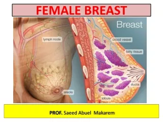
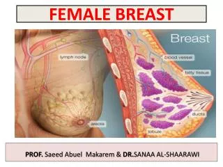
Parts, Shape & position of the Gland • It is conical in shape. • It lies in superficial fascia of the front of chest. • It has a base, apex and tail. • Its base extends from 2nd to 6th ribs. • It extends from the sternum to the midaxillary line laterally. • It has no capsule.
SHAPE AND POSITION OF FEMALE BREAST • 2/3 of its base lies on the pectoralis major muscle, while its inferolateral 1/3 lies on: • Serratus anterior & • External oblique muscles. • Its superolateral part sends a process into the axilla called the axillary tail or axillary process.
SHAPE AND POSITION OF FEMALE BREAST • Nipple: • It is a conical eminence that projects forwards from the anterior surface of the breast. • The nipple lies opposite 4th intercostal space. • It carries 15-20 narrow pores of thelactiferous ducts. • Areola : • It is a dark pink brownish circular area of skin that surrounds the nipple. • The subcutaneous tissues of nipple & areola are devoid of fat.
STRUCTURE OF MAMMARY GLAND • It is non capsulated gland. • It consists of lobes and lobules which are embedded in the subcutaneous fatty tissue of superficial fascia. • It has fibrous strands (ligaments of cooper) which connect the skin with deep fascia of pectoralis major. • It is separated from the deep fascia covering the underlying muscles by a layer of loose areolar tissue which forms the retromammary space.? What is its Importance? (allows the breast to move freely).
STRUCTURE OF MAMMARY GLAND • It is formed of 15-20 lobes. • Each lobe is formed of a number of lobules. • The lobes and lobules are separated by interlobar and interlobular fibrous & fatty tissue, called ligaments of Cooper. (Importance?) • It has from 15-20 lactiferous ductswhich open by the same number of openings on the summit of the nipple.
ARTERIAL SUPPLY • 1. Perforating branches of internal thoracic (internal mammary) artery. • 2. Mammary branches of lateral thoracic artery. • 3. Mammary branches ofIntercostal arteries.
VENOUS SUPPLY • Veins are corresponding to the arteries. • Circular venous plexus are found at the base of nipple. • Finally, veins of this plexus drain into axillary & internal thoracic veins.
AXILLARY LYMPH NODES • They are arranged into 5 groupswhich lie in axillary fat : • Pectoral (Anterior) group : which lies on the pectoralis minor along lateral thoracic vessels. • Subscapular (Posterior) group : which lies on posterior wall of axilla on lower border of subscapularis along subscapular vessels. • Brachial (Lateral) group : lies on lateralwall of axilla along 3rd part of axillary vessels. • Central group : lies in axillary fat at the base of axilla. • Apical group : lies at apex of axilla. • Subclavian lymph trunk: • It is formed by union of efferent lymph vessels of apical group. It usually opens in subclavian vein. On the left side it usually opens into thoracic duct.
LYMPHATIC DRAINAGE • Subareolar lymphatic plexus : • Lies beneath the areola. • Deep lymphatic plexus: • Lies on the deep fascia covering pectoralis major. • Both plexusesradiate in many directions and drain into different lymph nodes.
LYMPHATIC DRAINAGE • Central & lateral parts of the gland (75%) drain into pectoral groupofaxillary lymph nodes. • Upper part of the gland drains into apical groupof axillary lymph nodes. • Medial part drains into internal thoracic (parasternal) lymph nodes, forming a chain along the internal thoracic vessels. • Some lymphatics from the medial part of the gland pass across the front of sternum to anastomose with that of opposite side. • Lymphatics from the inferomedial part anastomose with lymphatics of rectus sheath & linea alba, and some vessels pass deeply to anastomose with the sub diaphragmatic lymphatics.
APPLIED ANATOMY- CANCER BREAST • It is a common surgical condition. • 60% of carcinomas of breast occur in the upper lateral quadrant. • 75% of lymph from the breast drains into the axillary lymph nodes. • In case of carcinoma of one breast, the other breast and the opposite axillary lymph nodes are affected because of the anastomosing lymphatics between both breasts. • In patients with localized cancer breast,a simple mastectomy, followed by radiotherapy to the axillary lymph nodes is the treatment of choice.
Applied Anatomy • The lactiferous ducts are radially arranged from the nipple,so incision of the gland should be made in a radial direction to avoid cutting through the ducts. • Infiltration of the ligaments of Cooper by breast cancer leads to its shortening giving peaude’orangeappearance of the breast.
Mammary ridge • Mammary ridge extends from the axilla to the inguinal region. • In human, the ridge disappears EXCEPT for a small part in the pectoral region. • In animals, several mammary glands are formed along this ridge.
Which is correct regarding the mammary gland ? It extends from the 2nd to 8th ribs. Its base lies on the pectoralis major muscle. It has 4-8 lactiferous ducts. Its most lymph drains into the parasternal lymph nodes. The lymphatics from upper part of mammary gland drain into : The parasternal lymph nodes. Subdiaphragmatic lymph nodes. Apical group of axillary lymph nodes. Pectoral group of axillary lymph nodes. The lactiferous ducts of mammary gland are : Less than 10. From 10-15. From 15-20. More than 20.