Download
1 / 25
270 likes | 1.05k Views
Seronegative Spondyloarthropathies Internal Medicine/Pediatrics Noon conference series June 1, 2006. Axial skeleton Skull Vertebral column Vertebrae Sacrum Coccyx Ribs Sternum Appendicular skeleton Girdles Extremities. The skeleton. Back to basics. Diarthrosis (moveable)

E N D
Seronegative SpondyloarthropathiesInternal Medicine/PediatricsNoon conference seriesJune 1, 2006
Axial skeleton Skull Vertebral column Vertebrae Sacrum Coccyx Ribs Sternum Appendicular skeleton Girdles Extremities The skeleton Back to basics
Diarthrosis (moveable) Majority of articulations Contiguous bones are covered by cartilage, connected by ligaments, and have an interposing synovial sac Synarthrosis (immoveable) Contiguous bones are in direct contact without cartilage, syovium, or ligaments Amphiarthrosis (sort of moveable) Characteristics of both diarthrosis and synarthrosis Contiguous surfaces are either: Connected by fibrocartiganeous disks (vertebral joint) Covered by fibrocartilage and partial synovium, and attached by external ligaments (sacroiliac joint) Articulations Back to basics
Enthesis is the site of bony attachment of Tendon Ligament Cartilage Joint capsule Fascia Enthesis Back to basics
Ankylosing spondylitis (the prototype) Psoriatic arthritis Reactive arthritis Formerly called Reiter’s syndrome) Enteropathic arthritis Undifferentiated spondyloarthropathy Mnemonic is PURE-A (sort of like purée) Comprise these conditions… Seronegative spondyloarthropathies
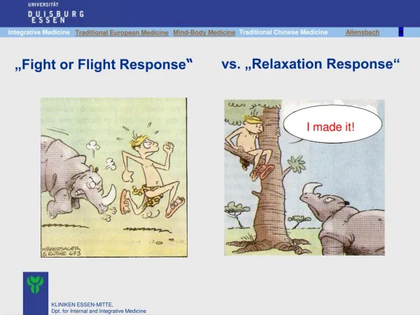
HLA-B27 association Enthesitis (both juxtaärticular and extraärticular) Axial skeleton arthritis (generally secondary to juxtaärticular enthesitis) Spondylitis (inflammation of vertebral bodies) Sacroiliitis (inflammation of sacroiliac joint) Peripheral arthritis (generally a synovitis) Asymmetric (cf rheumatoid arthritis) Extraärticular manifestations (besides enthesitis) Seronegativity Rheumatoid factor and ANA negative Well, because they share these characteristics… Why are these diseases classified together?
Ankylosing spondylitis: 95% Ethnically matched controls: 8% Reactive arthritis: 70% Enteropathic arthritis: 50% Psoriatic arthritis: 35% HLA-B27 association Why are these diseases classified together?
Inflammation of an enthesis Principal pathogenetic mechanism in spondyloarthropathy Pathogenesis CD8 T cells infiltrate entheses Activated macrophages release cytokines (egTNF) Fibroblasts synthesize new collagen (cf rhematoid arthritis!!) New bone formation results Clinical Axial skeleton arthritis (see later) Enthesopathy at other sites Calcaneal spurs at plantar fascia insertion Spurs at Achilles tendon insertion Manifests as extraärticular or juxtaärticular bony tenderness Enthesitis Why are these diseases classified together?
Arises from enthesitis Includes spondylitis and sacroiliitis Spondylitis CD8 T cells invade the junction of the annulus fibrosis and the vertebral body (an enthesis) Annulus fibrosis is replaced by bone (syndesmophytosis) Vertebral bodies assume a square shape, and ultimately a bamboo spine Sacroiliitis CD8 T cells invades the subchondral area at the junction of the bones and the cartilage (an enthesis) Cartilage on iliac side is replaced by bone, obliterating the jont space and hardening the joint Axial skeleton arthritis Why are these diseases classified together?
Inflammatory back pain requires 4 of these 5 criteria (serves as a screening tool for AS) Young onset ( 40 years) Morning stiffness ( 30 minutes) Chronic ( 3 months) Activity improves the pain (rest does not) Insidious (not acute) (mnemonic is YMCA-I) Diffuse lumbar or gluteal, not focal or radicular Cf focal pain of disk herniation Inflammatory back pain Ankylosing spondylitis
Restriction of lumbar movement Shober’s test – mark the patient’s back at the level of the posterior iliac spine. Place one finger 5 cm below this mark and a 2nd finger 10 cm above this mark. Patient is instructed to touch his toes. If the distance between finegrs increases < 5 cm, lumbar flexion is limited. Anterior uveitis (iritis or iridocyclitis) (25%) Acute eye pain Increased lacrimation Photophobia Blurred vision Aortitis with fibrosis Aortic insufficiency Third degree heart block (5%) Other clinical (besides back pain) Ankylosing spondylitis
Ankylosing spondylitis Radiographic evaluation
Low back pain 3 months improved by exercise and not relieved by rest Limitation of lumbar spine in sagittal and frontal planes Chest expansion reduction relative to normal values corrected for age and sex (costovertebral ankylosis, 25%) Radiographic criteria of sacroiliitis Bilateral grade 2-4 OR Unilateral grade 3-4 Ankylosing spondylitis is defined by the presence of either radiographic criterion PLUS any clinical criterion Modified New York Diagnostic Criteria Ankylosing spondylitis
In 1916, Hans Reiter reported Reiter’s syndrome: a triad of nongonococcal urethritis, conjunctivitis, and arthritis that occurred in a young German officer following an episode of bloody dysentery Subseqently, more cases were reported following enteric infections OR venereally acquired genitourinary infections. In 1967, the term reactive arthritis was applied to similar cases following Yersinia gastroenteritis The two terms should be considered synonomous The term reactive arthritis is increasingly preferred Interesting historical backdrop Reactive arthritis
Clinical syndrome triggered by specific etiologic agents in a genetically susceptible host Follows 1-4 weeks after a Urogenital infection (affects principally men) Usually C. trachomatis Enteric infection (affects both genddrs equally) Salmonella Shigella Campylobacter Yersinia Pathogenesis Reactive arthritis
Peripheral arthritis Asymmetric additive oligoarthritis (usually) Synovitis Warm Edematous Tender Pain with active or passive movement Usually lower extremity joints (knee, ankle, subtalar) Conjunctivitis Clinical Reactive arthritis
Nongonococcal urethritis Occurs in postenteric or postvenereal disease When it occurs in postvenereal disease, C. trachomatis is often the etiology When present, is usally the first symptom In men Mild dysuria Mucopurulent urethral discharge May present as prostatitis or epididymitis In women Dysuria Purulent vaginitis or cervicitis with vaginal discharge Asymptomatic urethritis often features sterile pyuria Clinical Reactive arthritis
Keratoderma blenorrhagica A papulosquamous skin rash Comprises vesicles that become hyperkeratotic, forming crusts before disappearing Palms/soles Penis (causing circinate balanitis Oral ulcers (ususally shallow and painless) Inflammatory back pain (50% of patients) Enthesitis (40%) Dactylitis (40%) Anterior uveitis (20% of patients) Clinical (continued) Reactive arthritis
Keratoderma blenorrhagica Reactive arthritis
Synovial fluid analysis Pleocytosis (5 000 to 50 000 WBC/mcL) with polymorphonuclear cell predominance Protein levels Glucose normal Cf reduced glucose level in true septic arthritis Gram stain and culture are sterile Urethral or cervical smears in patients with clinical urethritis C. trachomatis N. gonorrhoeae Evaluation Reactive arthritis
Affects 10-20% of patients with inflammatory bowel disease (IBD) Peripheral arthritis affects 10-20% of IBD patients Generally affects knees, ankles, and feet Always indicates active IBD Radiographic axial arthritis affects 10% of IBD patients Frequently asymptomatic Independent of bowel inflammation Clinical Enteropathic Arthritis
Nonsteroidal antiinflammatory agents Indamethacin Disease modifying anti-rheumatic drugs (DMARDs) Methotrexate: inhibits recruitment of CD4 and CD8 T cells Tumor necrosis factor antagonists Infliximab: a monoclonal antibody that binds to TNF and inhibits binding of TNF to its receptor Etanercept: similar emchanism to infliximab For axial arthritis, exercises to maintain posture and flexibility Treatment Why are these diseases classified together?