Download
1 / 1
10 likes | 165 Views
Blood pressure relaxing mechanisms in human endothelial cells after vertical stress. FLI. Shamci Monajembashi, Karl Otto Greulich. erythrocyte. Phase Contrast. The laser technique: Simulation of blood pressure by EMFA ( E rythrocyte M ediated F orce A pplication).

E N D
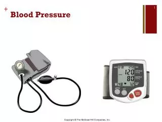
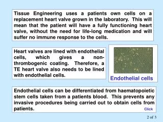
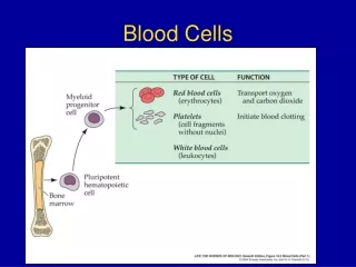
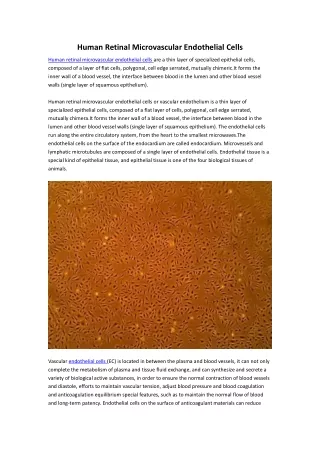
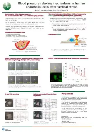
Blood pressure relaxing mechanisms in human endothelial cells after vertical stress FLI Shamci Monajembashi, Karl Otto Greulich erythrocyte Phase Contrast The laser technique: Simulation of blood pressure by EMFA (Erythrocyte Mediated Force Application) Hypertension (high blood pressure): The endemic disease No.1 and a normal aging process • EMFA technique exerting perpendicular forces to endothelial cells simulates blood pressure on single cell level. Application of EMFA in in vitro experiments may help: • To understand molecular and physiological mechanisms of blood vessel tone regulation. • To simulate the reaction of the vascular system to changes in blood pressure and thus to test new drugs in vitro at the level of endothelial cells. Understanding signal transduction in blood pressure research is the basis for new drugs. So far, principally shear stress and cyclic stretch are used for studies on the response of cells to hemodynamic forces. However, it is very little reported about response of endothelial cells to high vertical pressure [specially, in short time scale (a few seconds)]. Calcium Hemodynamic forces in vivo Principle of EMFA • Shear stress due to blood flow • Hydrostatic pressure due to blood pressure • Cyclic stretch due to deformation of vessel wall Fixed erythrocyte which is attached on the surface of endothelial cell, acts as a handle for optical tweezers to deform the cell HUVEC (Human Umbilical VeinEndothelial)Cells react by dramatic morphology changes after pressurizing HUVEC cells become stiffer after prolonged pressurizing Laser power: 460 mW for 6 sec;Total observation time: 27 min. Cell retraction changed after 6 min and lasted for 6 min (B-C). Cell recovered after 18 min (D). With slowly increased blood pressure (e.g. with age), cells can adapt themselves the high pressure. Rapidly increasing the blood pressure (e.g. from 120/80 mmHg to 180/100 mmHg) can not be any more adapted by the endothelial cells and they react dramatically to the high pressure increase. Laser power: 460 mW for 6 sec; Total observation time: 32 min. Relaxation started 70 sec after EMFA pressure and lasted for 30 min (B-C); Viable cell after 24h (D). • Perspectives • EMFA is the only experimental method which stimulates endothelial cells in true accordance to the blood pressure characteristics. • The combined mechanical and physiological response of endothelial cells to EMFA offers new possibilities in diagnosis and therapy of diseases based on endothelial dysfunctions. • Such simultaneously observations of two complex physiological phenomena after precise physical influence are so far in the aging research without comparison. • Cooperation: • Götz Pilarczyk; KIP Uni Heidelberg, INF 227, 69120 Heidelberg • Technical help: • Gabi Günther, Silke Schulz Cell layers react differently from single cell Ca and NO correlate Ca2+oscillation induces a NO concentration burst Intercellular Interactions: Ca/NO Homeostasis after EMFA Pressure on one cell is transmitted to neighbouring cells Coupled Ca2+ and NO oscillation