Download

1 / 59
590 likes | 865 Views
Skin & its Appendages. Chapter 6. Integumentary System. Skin and its appendages (attachments) Body’s thinnest, largest, most important organ Appendages = hair, nails, skin glands Integument = “skin” Relatively flat organ; classified as cutaneous membrane. Structure of the Skin.

E N D
Skin & its Appendages Chapter 6
Integumentary System • Skin and its appendages (attachments) • Body’s thinnest, largest, most important organ • Appendages = hair, nails, skin glands • Integument = “skin” • Relatively flat organ; classified as cutaneous membrane
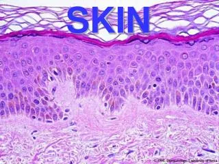
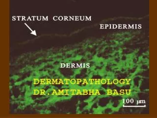
Structure of the Skin • Two main layers: • Outer, thinner layer (or strata) – epidermis • Composed of epithelial tissue • Inner, thicker layer of vascular connective tissue – dermis • Dermal-epidermal junction – where epidermis and dermis meet • Hypodermis – lies beneath the dermis (subcutaneous tissue) • Rich in fat and areolar tissue • Fat content varies with state of nutrition
Structure of the Skin • Thin and thick skin – refers to epidermis ONLY • Thin skin – covers most of body surface • Thick skin – palms of hands, soles of feet, finger tips • All 5 layers of epidermis present • Raised dermal papillae (fingerprints or footprints) • No hair is present in thick skin
Epidermis – Cell Types 3 cell types • Keratinocytes: synthesize keratin • Keratin: tough, fibrous protein found in hair, nails and outer skin • Keratinocytes make up 90% of epidermal cells • Principal structural element of outer skin • Melanocytes: synthesize melanin (brown pigment – gives skin color) • Melanin: protects deeper layers from ultraviolet light • Langerhan cells (immune cells) • Provide defense mechanism for the body • Arise from bone marrow & migrate to the epidermis
Epidermis – Cell Layers 5 strata (cell layers) • Stratum corneum (horny layer) • Most superficial layer • Shingle-like dead cells • Cell shape = squamous • Cytoplasm replaced by keratin • Keratinization – process of cells formed from deeper layers, fill with keratin and push to the surface
Epidermis – Cell Layers 2. Stratum lucidum (clear layer) • Keratinocytes are anucleated and clear • Cytoplasm filled with Eleidin – a protein-bound lipid that will eventually turn into keratin • Blocks water penetration and loss • Present only in thick skin
Epidermis – Cell Layers • Stratum Granulosum (granular layer) • Keratinization begins in this layer • 2-4 layers of flattened cells • Cells in this strata are filled with granules called keratohyalin • Stratum spinosum (spiny layer) • 8-10 layers of irregular shaped cells with prominent intercellular bridges (connections) • Cells are rich with RNA for protein synthesis required for the production of keratin
Epidermis – Cell Layers • Stratum basale (base layer) • Single layer of columnar cells • Mitosis only occurs here • Cells originate from here migrate to superficial layers shed Stratum germinativum (growth layer) • Used to describe the stratum spinosum and the stratum basale together
Epidermal Growth & Repair • Regeneration time – time period required for population of cells to mature & reproduce • Time for new cell formation = rate of old keratinized cells flaking off • Helps maintain constant thickness of epidermis • Cells push upward into each layer die become keratinized desquamate (fall away/shed) • Regeneration time is approximately 35 days
Dermal-Epidermal Junction • Composed of basement membrane • Also contains specialized fibrous elements & polysaccharide gel that cements epidermis to dermis • Partial barrier to passage of some cells and large molecules
Dermis or corneum (“true skin”) • Thicker than epidermis • Protective function against mechanical injury • Storage area for water and electrolytes • Contains somatic sensory receptors (nerves & nerve endings) • Process information such as: pain, pressure, touch, temperature • Muscle fibers, hair follicles, sweat & sebaceous glands, blood vessels • Rich vascular supply of the dermis plays a critical role in regulation of body temperature
Papillary Layer of Dermis • Thin layer • Loose connective tissue with elastic and collagenous fibers • Dermal papillae – bumps that project into epidermis • Creates distinct ridges on epidermal surface of fingers & toes (finger/footprints); unique for every person • Creates a gripping surface
Reticular Layer of Dermis • Thick layer • Dense irregular connective tissue • Network of fibers – collagenous & elastic • Contains muscle – skeletal & smooth (allows for point of attachment for movement) • Skeletal muscle – scalp movement & facial expressions • Arrector pili muscles: small bundles of smooth muscle that causes hair to “stand on end” – which causes the skin around it to raise = “goosebumps” • Somatic sensory receptors for pain, pressure, touch and temperature • Ex: tactile (Meissner) corpuscle senses light touch & pressure
Dermal Growth & Repair • Does not shed & regenerate like the epidermis • Rapid regeneration only occurs during wound healing • Dense fibrous connective tissue fills wound/incision/injury site to form a scar • Langer’s Cleavage lines: collagenous fibers in dermal layer tend to orient themselves in patterns that differ from one body part to another
Dermal Growth & Repair • Stretch marks – due to dermal tearing heal results in tiny scars • Ex: pregnancy, rapid weight gain/growth
Questions – Answer in complete sentences • Identify the two main or primary layers of skin. What tissue type dominates each layer? • The terms thin and thick refer to which primary layer of skin? How do thick and thin skin differ? • Identify the three cell types found in the epidermis. Give a description of all three. • List the five layers (or strata) of the epidermis. Give a brief description of each layer. Is each layer found in both thin and thick skin? • What is the name of the glue-like layer separating the dermis from the epidermis? • What structure forms the bumps that produce ridges on the palms and soles? Which layer of the dermis is this structure located in? • Which layer is vascular: the epidermis or dermis? What important role does the dermal vasculature play?
What Determines Our Skin Color? Skin Pigments – 2 types • Melanin – brown pigment • Skin color determined by quantity of melanin • Melanocytes convert the amino acid tyrosine to melanin; process catalyzed with tyrosinase • Majority of melanocytes found in stratum basale • Amount of melanin produced depends on genetics, sun exposure, age and hormones ACTH) • Ex: Albinism (albino individuals) – tryrosinase is absent from birth; melanin cannot be synthesized
Skin Color • Carotene – yellow pigment • Pigment found in food • Can deposit in stratum corneum of thick skin (palms & soles of feet) Other determinants of skin color: • Changes in the vasculature • Blood vessels constrict = pale • Blood vessels dilate = blush • Poorly oxygenated hemoglobin (low oxygen saturation) causes cyanosis (bluish color to skin)
Functions of the Skin • Protection • Sensation • Movement without injury • Excretion • Endocrine functions (vitamin D) • Immunity • Temperature regulation
Protection • Surface film – thin film of emulsified materials spread over the skin’s surface • Produced from the mixture of residue/secretions from sweat & sebaceous glands with epithelial cells constantly being shed • Function of the surface film: • Antibacterial & antifungal activity, lubrication, hydration of the skin surface, buffering of caustic irritants, blockage of toxic agents • Keratin protects against dehydration, microorganisms, chemical/mechanical damage • Melanin – protects against ultraviolet radiation
Sensation • Sensory receptors serve as antennas that detect stimuli • Pressure, pain, temperature, touch • Ex: tactile corpuscle
Movement & Growth • Movement and growth of the body as a whole can occur due to the presence of elastic fibers in the skin
Excretion • Body regulates volume and chemical composition of sweat through functions of the skin • Skin influences fluid volumes in the body & waste products excreted from the skin • Ex: uric acid, ammonia, urea
Vitamin D Production • Endocrine process • First steps of vitamin D production occurs in the skin when exposed to ultraviolet light • Vitamin D synthesis is completed in the kidneys & liver
Immunity • Specialized cells are present in the skin • Ex: langerhan cells with help from helper T cells trigger immune reactions
Regulation of Body Temperature • Majority of heat production is the result of food metabolism and activity of muscles and glands (esp the liver) • Vasocontriction: prevents heat loss warm blood circulating deeper within the body • Vasodilation: increases heat loss increases skin’s blood supply heat lost to the external environment • Evaporation, radiation, conduction and convection • Homeostasis of body temperature controlled by negative feedback mechanism
Burns • Tissue injury or skin cell death that results from heat/fire, overexposure to ultraviolet light, contact with electrical current, corrosive chemicals
Burns – Estimating Affected Body Surface • Treatment & prognosis of a burn depends largely on the amount of skin surface affected • “Rule of palms” – the size of the patient’s palm is approximately 1% of their body surface • “Rule of nines” • Body surface divided into 11 areas (anterior and posterior) of 9% • Perineum (genital area) accounts for 1 %
Severity of Burns • First degree burns – usually caused by a sunburn • only epidermis is damaged • skin is red and swollen, no blistering • Second degree burns • Epidermis and dermis are damaged • damage to sweat glands, hair follicles, and sebaceous glands • Blisters, severe pain, swelling and scarring are common • Third degree burns (full thickness burns) • destruction of epidermis and dermis • tissue death extends below sweat glands and hair follicles • burning may involve fascia, muscle, and bone • Patient may be insensitive to pain immediately after due to destruction of nerve endings • Severe scarring
Hair • Hairless areas: palms, soles of feet, lips, nipples, some parts of genitalia • At approximately 6 months gestation – fetus is covered with lanugo • After birth vellus hair replaces lanugo • At puberty – terminal hair replaces vellus hair
Hair Growth • Growth begins when epidermal cells spread down into dermis to form a follicle • Follicle consists of 2 layers • Outer dermal root sheath • Epithelial root sheath (external & internal parts) • Follicle’s inner layer formed by stratum germinativum • Germinal matrix: a cap-shaped cluster of cells at the bottom of the follicle • It is the cells of the germinal matrix that are responsible for forming hair/hair growth
Skin Glands Sweat glands - most common • Two types: Eccrine & apocrine • Based on location, secretion & nervous system connections • Eccrine sweat glands - most numerous and wide spread • simple, coiled, tubular gland • Produces sweat or perspiration - a watery liquid rich in salts, ammonia, uric acid, urea and other wastes • Sweat also plays an important role in maintaining constant body temperature • Majority found on soles of feet, palms, forehead and upper torso 2. Apocrine sweat glands - larger than eccrine glands • located deep in the subcutaneous layer of the skin • Ex: armpit (axilla), areola of breast, pigmented skin around anus • Ducts are connected to and secrete into hair follicles • Classified as simple, branched tubular glands • Function of apocrine glands begins at puberty • Odor is from skin bacteria, not secretions
Sebaceous Glands • Secrete oil for hair & skin • Simple alveolar gland; found in dermal layer • Oil = sebum • Lubricates skin • Antifungal property – kills fungus & bacterial • Increases effectiveness of surface film • Mostly associated with hair follicles • Glands activated at puberty • Stimulated by sex hormones • Accumulation of sebum in ducts = white pimple • Oxidation causes sebum to darken = blackhead
Ceruminous Glands • Modified apocrine sweat gland • Open into the skin of the external ear cannal • Brown waxy substance = cerumen • Provides protection from dehydration • Risk for blockage and hearing loss
Mechanisms of Disease – Skin Disorders • Skin Infections • Vascular & Inflammatory Disorders • Abnormal Body Temperature • Skin Cancers
Skin Infections • Impetigo • Bacterial infection caused by either staphylococcus or streptococcus that usually occurs in young children • Reddish discoloration that develops into vesicles (blisters) and yellowish crusts • Can become systemic • Tinea – fungal infection (ex: ringworm, jock itch, athlete’s foot) • s/sx: erythema (redness), scaling, crusting • Tx: antifungal medications