Jaringan Otot
STRUKTUR HEWAN BAB 5. JARINGAN OTOT TIM DOSEN STRUKTUR HEWAN JURUSAN BIOLOGI FAKULTAS MATEMATIKA DAN ILMU PENGETAHUAN ALAM UNIVERSITAS PADJADJARAN. Jaringan Otot. Berfungsi dalam pergerakan tubuh

Jaringan Otot
E N D
Presentation Transcript
STRUKTUR HEWANBAB 5.JARINGAN OTOT TIM DOSEN STRUKTUR HEWANJURUSAN BIOLOGI FAKULTAS MATEMATIKA DAN ILMU PENGETAHUAN ALAM UNIVERSITAS PADJADJARAN
JaringanOtot • Berfungsidalampergerakantubuh • Dibangunolehsel-sel yang kayaakanmikrofilamensehinggadapatmelakukankontraksidanrelaksasi
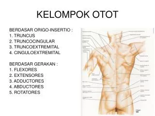
Berdasarkansifatmorfologi (struktur) danfungsinyaterdapattigajenisotot : Ototrangka, ototjantungdanototpolos • Tigajenisjaringanototdapatdibedakanpadamammaliaberdasarkancirimorfologisdanfungsional, dansetiapjaringanototmempunyaistruktur yang sesuaidenganperananfisiologisnya. • Ototrangkatersebarpadarangka, ototjantungterdapatpadajantung, danototpolostersebarluaspadasistemkardiovaskuler, pencernaanmakanan, urogenitaldanpernafasan.
Figure 10–1.Struktur of the 3 muscle types. The drawings at right show these muscles in cross section. Skeletal muscle is composed of large, elongated, multinucleated fibers. Cardiac muscle is composed of irregular branched cells bound together longitudinally by intercalated disks. Smooth muscle is an agglomerate of fusiform cells. The density of the packing between the cells depends on the amount of extracellular connective tissue present. Kontraksikuat, volunter, cepatdantidaksinambung Kontraksikuat, involunter, cepatdansinambung Kontraksilemah, involunter, lambat
OtotRangka Structure of the skeletal muscle. The drawing a show the muscle in cross section. Skeletal muscle is composed of large, elongated, multinucleated fibers. • Dibangunolehberkas-berkasserabutotot yang berintibanyakdanmenggambarkangaris-garismelintang • Intinyabanyakterletakdibagiantepiserabutotot • Kontraksikuat, volunter, cepatdantidaksinambung (terputus)
Figure 10–8. Longitudinal section of skeletal muscle fibers. Note the dark-stained A bands and the light-stained I bands, which are crossed by Z lines. Giemsa stain. High magnification OtotRangka • Serabutototterdiridarimiofibril-miofibril • Miofibrilterdiridari sub unit struktural yang disebutsarkomer - yang didalamnyaterdapatsusunan yang teraturdarifilamen-filamentebal (miosin) danfilamen-filamentipis (aktin). • Membran plasma serabutototdisebutsarkolemadanretikulumendoplasmiknyadisebutretikulumsarkoplasmik . Figure 10–6. Striated skeletal muscle in longitudinal section (lower) and in cross section (upper). The nuclei can be seen in the periphery of the cell, just under the cell membrane, particularly in the cross sections of these striated fibers. H&E stain. Medium magnification.
Figure 10–11. Structure and position of the thick and thin filaments in the sarcomere. The molecular structure of these components is shown at right. (Drawing by Sylvia Colard Keene. Reproduced, with permission, from Bloom W, Fawcett DW: A Textbook of Histology, 9th ed, Saunders, 1968.) • Strukturdanposisifilamentebaldantipispadasarkomer. • Sarkomermemperlihatkangambaranpolapita gelapdanterang. • Pita utamaadalahgelap (pita A) ditempatiolehfilamentebalsecarautuhdanolehsebagianfilamentipis, dan yang terang(pita I) yang hanyaberisikanfilamentipis. Filamen-filamentsbtersusunsejajarmenurutkepanjangansarkomer. • Satuujungfilamentipismelekatpadagaris Z. Di tengah-tengah pita A terdapatpita Hyghanyaberisikanfilamentebal. Bilaototmengkerut, makafilamen-filamentsbakan “sliding past another”
Membran plasma serabutototdisebutsarkolemadanretikulumendoplasmiknyadisebutretikulumsarkoplasmik . • RetikulumsarkoplasmikadalahmodifikasidariRetikulumendoplasmik, merupakanbagian integral darimekanisme yang mengaturkonsentrasikalsiumdisekelilingmiofibril. Retikulumsarkoplasmikmengelilingiberkas-berkasmiofibril. • DisampingRetikulumsarkoplasmik, terdapat pula sistemTubulus transversal. • Sisteminimerupakaninvaginasisepertijaridarisarkolemapadaketinggianpertemuan pita I dan pita A dalamsuatusarkomer, untukselanjutnyamembentuksistemtubulus yang bercabang-cabangdanberanastomose. Dengandemikiansatusarkomerdilayaniolehduasistemtubulus (T tubule). • Satutubulus T akanberhubungandenganduasisterna terminal membangunsuatu triad.
Serabutototrangkadiselaputiolehendomisium,suatujaringanikatlonggar yang terdiridarifibroblasdanserabutkolagen. • Serabutototmembentukberkasserabutotot /fasikulumototdiselaputiolehjaringanikat yang disebutperimisium • Sejumlahfasikulumototmembangunotot (misalnyaotot biceps) , yang diselaputiolehjaringanikat yang disebutepimisium Figure 10–3. Cross section of striated muscle stained to show collagens type I and III and cell nuclei. The endomysium is indicated by arrowheads and the perimysium by arrows. At left is a piece of epimysium. Picrosirius-hematoxylin stain. High magnification.
Figure 10–21. Section of tongue, an organ rich in striated skeletal muscle fibers. These fibers appear brown because the section was immunohistologically stained to show myoglobin. The light-colored areas among and above the muscle fibers contain connective tissue. In the upper region of the section, stratified and cornified epithelium can be seen. Nuclei are stained by hematoxylin. Low magnification. • Contoh : ototrangkapadalidah
OtotJantung • Selototjantungberbentukserabut yang bercabangdanberanastomosemembentukanyaman yang rapatdanmemperlihatkangaris-garismelintangpadaserabutnya • Mempunyaisatuintidalamsatuserabutototjantung • Pertemuanantaracabang-cabangserabutototjantungmembangunsuatuhubungan yang kompleks , disebutkeping (cakram) interkalaris. • Bekerjaritmis, terusmenerus, kuatdaninvolunter
Figure 10–26.Junctional specializations making up the intercalated disk. Fasciae (or zonulae) adherentes (A) in the transverse portions of the disk anchor actin filaments of the terminal sarcomeres to the plasmalemma. Maculae adherentes, or desmosomes (B), found primarily in the transverse portions of the disk, bind cells together, preventing their separation during contraction cycles. Gap junctions (C), restricted to longitudinal portions of the disk—the area subjected to the least stress—ionically couple cells and provide for the spread of contractile depolarization. • Terdapattigajenishubunganutama : fasia /zonulaadherens, makulaadherens (desmosom) dan gap junction (taut rekah) • Mengandungbanyakmitokondria yang menempati 40% ataulebihdari volume sitoplasma, danendomisiumnyakayaakanpembuluhdarah. • Strukturdanfungsidari protein kontraktildalamselototjantungpadadasarnyasamadenganototrangka, tetapisistemtubulus T danretikulumsarkoplasmiktidakbegituteratur. Tidakdijumpaiberkasmiofibril yang jelas. (taut rekah)
Potonganmemanjangbagiandariduaselototjantung. Bagiandiskusinterkalaris yang tersusunmelintangterdiriatasfasiaadherensdanbanyakdesmosom. Bagian yang memanjang (matapanah) mengandung taut rekah gap junction. Terdapatbanyakmitokondria (M). Tampakserat-seratretikulin (R) diantarakeduasel . 18.000 X Figure 10–23. Photomicrograph of cardiac muscle. Note the cross-striation and the intercalated disks (arrowheads). Pararosaniline–toluidine blue (PT) stain. High magnification
OTOT POLOS • Berbentuksepertigelendong/kumparanpanjangberukuran 30-200µm, sel-seltersusunrapat, berintisatuditengah, sitoplasmanyahomogen • Mengandungmiofilamenaktindanmiosin, tetapitidaktersusunteratursepertipadaototrangka, olehkarenaitutidakmenggambarkanseran-lintang. • Bekerjadiluarkehendak (involunter), kontraksilambat, tetapibekerjadalamwaktu yang lama • Terdapatpada organ-organ bagiandalam: Dindingsaluranpencernaan, kelenjarpencernaan, kantungurin, alatkelamin, saluranpernafasan, pembuluhdarah Figure 10–29. Photomicrographs of smooth muscle cells in cross section (upper) and in longitudinal section (lower). Note the centrally located nuclei. In many cells the nuclei were not included in the section. PT stain. Medium magnification.
OtotPolos Satusegmenototpolos, semuaseldikelilingijalinanseratretikulin. Seratretikulin (impregnasi Ag) pd sayatanmelintangototpolos, membentukjalinanygmengelilingisel-selototpolos • Selototpolosdibungkusoleh lamina basalisdanjalinanseratretikulin
Figure 17–9. Large bronchus. Note the distinct layer of smooth muscle that influences the flux of air in the respiratory system. PT stain. Medium magnification. • Ototpolospadasistemrespirasi
Figure 10–29. Photomicrographs of smooth muscle cells in cross section (upper) and in longitudinal section (lower). Note the centrally located nuclei. In many cells the nuclei were not included in the section. PT stain. Medium magnification.
Elektronmikrografpreparatsayatanmelintangototpolos, sertarelaksasi & kontraksiototpolos.
Figure 11–8. Cross section through an arteriole and its accompanying venule from the myometrium of mouse uterus. Note the elongated, large nucleus (arrowhead) of a pericyte surrounding the venule wall. Toluidine blue stain. High magnification. (Courtesy of TMT Zorn.) Contoh: Ototpolospadapembuluhdarah
Figure 10–33. Smooth muscle cells relaxed and contracted. Cytoplasmic filaments insert on dense bodies located in the cell membrane and deep in the cytoplasm. Contraction of these filaments decreases the size of the cell and promotes the contraction of the whole muscle. During the contraction the cell nucleus is deformed. • Selototpolosmemilikisuatujajaranfilamensepanjang 10 nm didalamsitoplasmanya. • Duajenisbadanpadatmunculdalamototpolos. • Filamensitoplasmiktsbmasukkedalambadanpadat yang berlokasipadaselmembrandansitoplasma. • Kontraksi filamen2 mengurangiukuranseldanmeneruskankekuatankontraksikepadasel-selototpolosberdekatandanseluruhotot.
RegenerasiOtot • Setelahterjadicedera, ketigajenisotot (dewasa) tsbmemilikipotensiregenerasiberbeda. • Ototjantunghampirtidakmempunyaipotensiregenerasisstelahmasaanak-anak, infark (kerusakan) digantikanolehproliferasijaringanikatdimiokardium. • Ototrangkaintiselnyatidakmampumelakukan mitosis, tapimempunyaiselsatelityang aktif (bilaadacedera) berproliferasidanbergabungmembentukserabutototbaru. • Ototpolosmampumemberiresponregenerasiaktif, bilaadacederaselototpolosmononukleusygmshhidupmengalami mitosis danmenggantikanjaringanygrusak