Download
1 / 53
570 likes | 1.08k Views
Blood Physiology Professor A.M.A Abdel Gader MD, PhD, FRCP (Lond., Edin), FRSH (London) Professor of Physiology , College of Medicine & King Khalid University Hospital King Saud University Riyadh. Hemostasis: the spontaneous arrest of bleeding from ruptured blood vessels Mechanisms:

E N D
Blood PhysiologyProfessor A.M.A Abdel GaderMD, PhD, FRCP (Lond., Edin), FRSH (London)Professor of Physiology, College of Medicine &King Khalid University HospitalKing Saud UniversityRiyadh
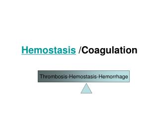
Hemostasis: • the spontaneous arrest of bleeding • from ruptured blood vessels • Mechanisms: • Vessel wall • Platelet • Blood coagulation • Fibrinolytic system
Memostatic Mechanisms: • Mechanisms: • Vessel wall • Platelet • Blood coagulation • Fibrinolytic system
Memostatic Mechanisms- cont • Vessel wall • Immediately After injury a localized Vasoconstriction • Mechanism -Hurmoral factors: • local release of thromboxane A2 & 5HT by platelets • Systemic release of adrenaline • Nervous factors
Platelets – cont. Site of formation: Bone marrow Steps: Stem cell Megakaryoblast Megakaryocyte Platelets
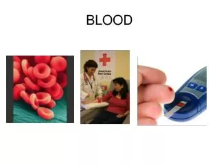
Platelets Thrombocytes are • Fragments of megakaryocytes in bone marrow
Platelets Formation (Thrombopoiesis) Regulation of thrombopoiesis by Thrombombopoietin
Platelet Functions Begins with Platelet activation
Platelet Activation • Adhesion • Shape change • Aggregation • Release • Clot Retraction
Platelet Adhesion • Platelets stick to exposed collagen underlying damaged endothelial cells in vessel wall
Platelet Release Reaction • Platelets activated by adhesion • Extend projections to make contact with each other • Release thromboxane A2, serotonin & ADP activating other platelets • Serotonin & thromboxane A2 are vasoconstrictors decreasing blood flow through the injured vessel. ADP causes stickiness & aggregation
Platelet Aggregation • Activated platelets stick together and activate new platelets to form a mass called a platelet plug • Plug reinforced by fibrin threads formed during clotting process
Platelet Activation • Adhesion • Shape change • Aggregation • Release • Clot Retraction
Platelet • Adhesion Endothelium 2. Shape change • 3. Aggregation
Platelet Plug Aggregation of platelets at the site of injury to stop bleeding • Exposed collagen attracts platelets • Activated platelets release of platelet ADP & TXA2the stickiness of platelets Platelets aggregation plugging of the cut vessel • Intact endothelium secret prostacyclin inhibit aggregation
Activated Platelets Secrets: • 5HT vasoconstriction • Platelet phospholipid (PF3) clot formation • Thromboxane A2 (TXA2) is a prostaglandin formed from arachidonic acid Function of TXA2: • vasoconstriction • Platelet aggregation (TXA2 inhibited by aspirin)
Extrinsic pathway Intrinsic Pathway Tissue factor FXII FXIIa Factor VII & Ca ++ FXI FXIa FIX FIXa FVIII X Xa FV+ Ca+P Prothrombin Thrombin Fibrinogen Fibrin The Coagulation Cascade
The “Cascade”, “Waterfall” model Main feature: Depicts the coagulation process as: a series of proteolytic reactions >> each protease cleaves and activates the subsequent protease
Activation Blood Coagulation • Intrinsic Pathway: all clotting factors present in the blood • Extrinsic Pathway: triggered by tissue factor • Common Pathway
Fibrinolysis Plasminogen activators Plasminogen Plasmin Anti-activators Anti-plasmin Coagulation Fibrin FDP The fibrinolytic System
ANTICOAGULANTS Anticoagulants: Substances that prevent blood coagulation
Anticoagulants-cont. Types: • Substances that remove Ca++ from the blood: Citrate, oxalate, EDTA (Sequestrene) Ethylene Diamine Tetra chloro-Acetate
Anti-coagulants-cont. 2. Heparin • Action: • inhibit the conversion of prothrombin to thrombin • inactivate thrombin (anti-thrombin III) • Uses: • Open heart surgery, Post-operative to prevent deep vein thrombosis • Collection of blood samples in labs.
Anticoagulants-cont. 3. Warfarin • Inhibits the synthesis of Vit-K dependent clotting Factors (II, VII, IX & X) by the liver • Widely used in medicine as a oral anticoagulant
Anticoagulants-cont. 4.Naturally occurring anticoagulants: • Control intravascular clotting • Antithrombin III inhibit thrombin, X, VIII, IX, XII • Protein C inhibit factor VIII, V
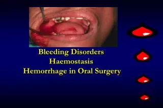
Bleeding Disorders Diseases characterized by excessive bleeding: • Vascular defects • Congenital • Acquired • Platelet defects • Low platelet count Thrombocytopenia 3. Coagulation defects • Congenital • Acquired
3. Coagulation defects • Congenital: • Haemophilia A(deficient factor VIII), • Haemophilia B (deficient factor IX, Chrismas Disease) Treatment: • Replacing the missing factor • Transfusing: fresh frozen plasma, clotting factor concentrate