Download
1 / 157
1.57k likes | 1.62k Views
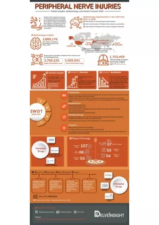
Explore the microscopic anatomy, internal topography, classification, etiology, and clinical diagnosis of peripheral nerve injuries. Learn about electrodiagnostic studies, diagnostic tests, factors influencing regeneration, nerve repair techniques, and brachial plexus injuries. Discover various treatment methods and factors affecting nerve regeneration post-neurorrhaphy.

E N D
PERIPHERAL NERVE INJURYIES MHD BASHAR ALBOSHI
NEURONAL DEGENERATION AND REGENERATION: • Phagocytosis • Secondary or wallerian degeneration • Primary or retrograde egeneration
CLASSIFICATION OF NERVE INJURIES: ( Seddon1943): • Neurapraxia • Axonotmesis • neurotmesis • (Sunderland 1951) classification
Etiology of peripheral nerve injuries: - Metabolic or collagen diseases - Malignancies -Endogenous or exogenous toxins -Thermal -Chemical -Mechanical trauma
Clinical diagnosis of nerve injuries: Highet Scale: 0 – total paralysis. 1- muscle flicker. 2-muscle contraction. 3- muscle contraction against gravity. 4- muscle contraction against gravity and resistance. 5-normal muscle contraction .
Diagnostic tests: Electrodiagnostic studies provide the clinician with a base of knowledge as follows:: 1-Documentation of injury Location of insult2 - 3-Severity of injury 4-Recovery pattern 5-Prognosis 6-Objective data for impairment documentation 7-Pathology 8-Selection of optimal muscles for tendon transfer 9-procedures
The most common electrodiagnostic methods used for the study of peripheral nerve injuries are : @- nerve conduction studies and @-electromyography (EMG)
Fig. 59-10 Diagram of EMG tracing depicting normal insertion activity, which also may be present immediately after denervation.
Fig. 59-11A, Diagram of EMG tracing demonstrating positive sharp wave consistent with denervation 10 to 14 days after injury. Rhythm is regular, amplitude is 100 to 400 uV, duration is 5 to 150 msec, and rate is 2 to 40 Hz. B, Diagram of EMG tracing demonstrating spontaneous denervation fibrillation potentials present within 14 to 18 days after injury. Rhythm is regular, amplitude is 50 to 1000 uV, duration is 0.5 to 2 msec, and rate is 2 to 30 Hz.
Tinel sign : A positive Tinel sign is presumptive evidence that regenerating axonal sprouts that have not obtained complete myelinization are progressing along the endoneurial tube. @- neuropraxia(sunderland1) -------negative Tinel sign. @- axonotmesis (sunderland2,3) -------positive Tinel sign. (sunderland4-------- negative Tinel sign ) @- neurotmesis (sunderland 5) ------- negative Tinel sign. Other diagnostic test: Sweat test.,skin resistance test, electrical stimulation
GENERAL CONSIDERATIONS OF TREATMENT. FACTORS THAT INFLUENCE REGENERATION AFTER NEURORRHAPHY : 1-Age 2-Gap Between Nerve Ends 3-Delay Between Time of Injury and Repair 4-Level of Injury5-Condition of Nerve Ends
TECHNIQUE OF NERVE REPAIR: Endoneurolysis (Internal Neurolysis Partial NeurorrhaphyNeurorrhaphy and Nerve Grafting
Methods of Closing Gaps Between Nerve Ends: Mobilization Positioning of Extremity Transposition Bone Resection Nerve Stretching and Bulb Suture Nerve Grafting
Techniques of Neurorrhaphy: Epineurial Neurorrhaphy
ETIOLOGY AND CLASSIFICATION OF BRACHIAL PLEXUS INJURIES : -birth, -missiles, - stab wounds, - traction applied to the plexus during falls, -vehicular accidents, - sports activities, -as well as radiation.
Rupture of the axillary or subclavian artery occurs in 20% of patients. Common associated injuries include: @fractures of the proximal humerus, the scapula, the ribs, the clavicle, and the transverse processes of the cervical vertebrae @dislocation of the shoulder, the acromioclavicular, and the sternoclavicular joints. @ A torn rotator cuff also has been described in conjunction with brachial plexus injury.
Upper plexus injury (Erb) involves the segments innervated by the C5 and C6 nerve roots with or without dysfunction of the C7 root. -Typically the limb is extended at the elbow(the biceps, brachialis, and brachioradialis muscles) -flaccid at the side of the trunk, • -Adducted (deltoid and supraspinatus muscles) • -internally rotated (infraspinatus and teres minor muscles, supinator muscle)
Lower plexus injury (Klumpke) can be diagnosed by finding segmental sensory and motor deficits involving C8 and T1 with or without C7 dysfunction. @ The primary dysfunction is apparent in : -the intrinsic musculature of the hand along with paralysis of the wrist and finger flexors. - The sensory deficit is along the medial aspect of the arm, forearm, and hand. @Associated Horner syndrome should alert the examiner to the possibility of an avulsing injury of the lower plexus, .
Injuries to the upper or lower trunks produce essentially the same sensory and motor deficits as do injuries to their respective rami, except for preservation of function of the long thoracic and dorsal scapular nerves in the upper trunks and absence of Horner syndrome in the lower trunks.
Injuries of the lateral cord deficits in the distribution of: • -musculocutaneous nerve (paralysis of the biceps). • -lateral root of the median nerve (paralysis of the flexor carpi radialis and pronator teres). • -lateral pectoral nerve (clavicular head of the pectoralis major). • @Glenohumeral subluxation may result. This may be prevented by an aggressive program of rehabilitation of the remaining intact musculature. • @Sensory deficit can be detected over the anterolateral aspect of the forearm in the relatively small autonomous zone of the musculocutaneous nerve.
Injuries of the posterior cord deficits in the distribution of the following nerves: -subscapular (paralysis of the subscapularis and teres major), -thoracodorsal (paralysis of the latissimus dorsi). - axillary (paralysis of the deltoid and teres minor), -radial nerve (paralysis of extension of the elbow, wrist, and fingers). @The disability consists mainly of inability to internally rotate the shoulder, elevate the limb, and extend the forearm and hand. @ Sensory loss most often is apparent only in the autonomous zone of the axillary nerve overlying the deltoid muscle..
Injuries of the medial cord produce the motor deficit of: - a combined ulnar and median nerve lesion (except for the flexor carpi radialis and pronator teres) and - extensive sensory loss along the medial aspect of the arm and hand
TREATMENT OF BRACHIAL PLEXUS INJURIES Open injuries: exploration and primary repair can be attempted. Usually, however, injuries to adjacent vessels or to the mediastinal or thoracic viscera must be treated first, and thus repair of the plexus injury must be delayed. @. Leffert emphasized the poor prognosis after lower trunk injuries but advised surgical exploration for sharp injuries of the upper and middle trunks. @When an open injury has been caused by a low-velocity missile, early exploration is not indicated unless injuries to adjacent vessels or viscera make immediate treatment necessary. @Consequently a period of observation is indicated because considerable function may return spontaneously @Again electromyograms should be obtained 3 to 4 weeks after injury to aid in determining the extent of denervation. Thereafter periodic examinations are indicated every 4 to 6 weeks @When such examinations during a reasonable period of time reveal the absence of recovery or that any recovery has halted, exploration and neurorrhaphy, grafting, or neurolysis may be beneficial
Surgical Goals Surgical GoalsThe surgeon should have clear and reasonable surgical goals, which are in order of priority: (1) restoration of elbow flexion. (2) restoration of shoulder abduction. (3) restoration of sensation to the medial border of the forearm and hand. @After brachial plexus repair and reconstruction, 12 to 18 months are required to determine the extent of neural regeneration. /
. Tendon transfers about the shoulder that may be considered include • trapezius to deltoid transfer to improve abduction • -latissimus dorsi transfer to improve external rotation. • - Shoulder arthrodesis is helpful if active scapulothoracic motion is preserved and has been shown to improve elbow flexion by preventing uncontrolled internal rotation of the shoulder. The shoulder should be fused in only 20 to 30 degrees of abduction because most of these patients..
@Operations to restore elbow flexion include transfers of the latissimus dorsi, the pectoralis major, the triceps, the sternocleidomastoid, and the flexor-pronator mass). - Marshall et al. reviewed 50 such transfers and found the latissimus dorsi and triceps transfers to be the most reliable. Restoration of elbow flexion is helpful to the patient even if the hand is functionless
The long thoracic nerve arises from C5, C6, and C7 immediately after they emerge from the intervertebral foramina.
. It traverses the neck posterior to the brachial plexus, continues distally along the lateral aspect of the thoracic wall, and innervates the serratus anterior muscle.
injuries may result from: • -either sharp or blunt trauma • - or from traction when the head is forced acutely away from the shoulder or when the shoulder is depressed, as when carrying heavy weights. • - Other causes include exposure to cold, viral infections, and placing patients in the Trendelenburg position with shoulder braces that compress the supraclavicular areas.