Head Segmentation using Finite Element Method Approach
10 likes | 108 Views
Detailed models of the cerebral cortex from MRI data are essential for brain studies. This paper introduces a new framework based on finite element method to segment head interfaces from anatomical MRI, enhancing accuracy and automation.

Head Segmentation using Finite Element Method Approach
E N D
Presentation Transcript
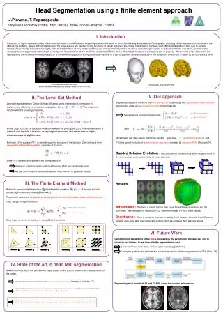
Head Segmentation using a finite element approach J.Piovano, T. Papadopoulo Odyssée Laboratory (ENPC, ENS, INRIA), INRIA, Sophia-Antipolis, France I. Introduction Extraction of highly detailed models of the cerebral cortex from MRI data is extremely useful in the study of brain functionality and anatomy. For example, accuracy of the segmentation is crucial in the MEG/EEG problem, where distinct interfaces of the head tissues are needed to find locations of neural activity in the cortex. Extraction of surfaces from MRI data is a difficult task due to several factors. Anatomically, the cortex is a highly convoluted thin layer of grey matter and because of the complexity of the structure, manual segmentation is tedious and often unfeasible, so automated computer-based segmentations methods are required. Segmentation of the skull from anatomical MRI is also a difficult task because it cannot be seen in properly . We present a new framework for representing and evolving level sets, based on a finite element approach and quadrilateral meshes, in order to segment several interfaces of the head from anatomical T1 (and T2 as future work) MRI. Example of an anatomical MRI, where unknown interfaces are already colored (from JP.Pons) Interfaces in 3D (from JP.Pons) V. Our approach Segmentation is done thanks to the level set method implemented with Quadrilateral finite elements and evolving under a regionbased evolution [Chan-Vese:99]. two equations solved simultaneously With represent the “step values” to find the function at time (implicit scheme), and is the speed evolution of a region based approach, modeled by Gaussian PDE [Rousson:04] Banded Scheme Evolution : we compute the solutions only at the neighborhood of the zero levelset, and maintain a list of active elements Results : Advantages: The band is clearly thinner than usual finite difference scheme, and the subvoxelic representation of the solution (Q1 elements instead of P1)) is more natural Drawbacks : Slow to compute, and gain in quality is not assured, as usual finite difference scheme work quite well, are robust, and grid on which we compute them are very dense. II. The Level Set Method Level set representations [Osher–Sethian:88] are a useful mathematical formulation for implementing efficiently curve/surface propagation. Let be a Lipschitz function with the following properties, where is the euclidean distance between the pixel and . This representation is intrinsic and implicit, it imposes no topological constraint and extensions to higher dimensions are straightforward. Evolution of the surface is done through the evolution of the function by solving a time-dependent differential equation, generally of the form: Where F is the evolution speed in the normal direction Numerical schemes based on finite differences ENO are traditionally used We can use numerical schemes based on finite element to get better results Evolution equation Eikonal equation III. The Finite Element Method Method to approximate the solution of a differential equation through a mesh partitioning the resolution space [Zienkewicz]. The solution values are computed at vertex positions, and interpolated inside each elements. Thus, we get the approximation, Many types of elements, leading to many different solutions . Solutions values at vertex locations Interpolation basis, « shape functions » VI. Future Work Using the high capabilities of the GPUs, to speed up the evolution of the level-set and to visualize and interact in real time with the segmentation result. Some work have been done, and are quite promising [Lefohn:03] Conjugate gradient (the bottleneck of our framework) have been programmed on GPU [Bolz : 02] Segmenting skull from both T1 and T2 MRI, using the coupled information. IV. State of the art in head MRI segmentation Several methods, each one with several steps, based on the a priori anatomically representation of the cortex. Denoising + bias removing + Tissus classification with Expectation-maximisation based methods [Dempster-Laird-Rubin : 77] Using the fact that the cortex is a layer of nearly constant thickness, and use a system of coupled surfaces initialized as two concentric spheres with an additional force which prevent them to move away, of be too close to each other.[Zeng : 99] Using a region based evolution speed, based on the fact that the head can be decomposed in 3 homogeneous regions: white matter, grey matter, CSF+skull (seen as black values), and let the evolution converge to retrieve 3 different regions, one of them being the cortex[Chan–Vese : 99]