Download
1 / 61
610 likes | 614 Views
Introduction to the Reproductive System. Sexual reproduction produces new individuals Gametes are sperm & egg Formed by testes and and ovaries Fertilization produces one cell (a zygote)with one set of chromosomes from each parent Creates genetic variation.

E N D
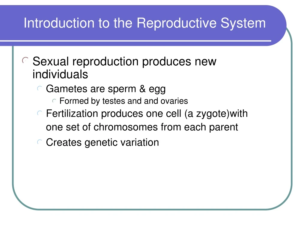
Introduction to the Reproductive System • Sexual reproduction produces new individuals • Gametes are sperm & egg • Formed by testes and and ovaries • Fertilization produces one cell (a zygote)with • one set of chromosomes from each parent • Creates genetic variation
Introduction to the Reproductive System • Gonads (testes & ovaries) produce gametes & secrete sex hormones • Reproductive systems • Gonads, ducts, glands & supporting structures • Gynecology is study of female reproductive system • Urology is study of urinary system & male reproductive system
Male Reproductive System Ejaculatory Duct- sperm and seminal fluid into urethra Seminal vesicle-seminal fluid Ductus deferens- transports sperm Prostate-semen Urethra-urine & semen Penis Epididymis-sperm maturation Testis- sperm formation Scrotum-cools testis
Testis • Contains 200 - 300 compartments called lobules • Each lobule contains 2 or 3 seminiferous tubules where sperm are formed • Sperm formation called spermatogenesis Lobule Seminiferous tubule
Seminiferous Tubules and Spermatogenesis • Seminiferous tubules contain • Sperm forming cells called spermatocytes that become sperm • Supporting cells called Sertoli cells support sperm and secrete substances important to sperm formation • Interstitial cells in between tubules secrete testosterone Sperm
Spermatogenesis • Spermatocytes in seminiferous tubules • Undergo meiosis • Results in four cells with 23 chromosomes • Produces new combinations of genes • Each spermatocyte forms four sperm ( spermatozoa) • Steps are shown on next slide, but may not be enough time to cover it in class
SpermatogenesisSperm forming cells go through two meiotic divisions 1°=primary 2°=secondary • Each of four cells develop into sperm • Second division results in four spermatids,each with 23 single stranded chromosomes • First division results in two 2° spermatocytes, each with 23 double stranded chromosomes • Starts with a 2n=46 1° spermatocyte
Sperm Structure • Adapted for reaching & fertilizing egg • Head contains DNA & enzymes for penetrating to egg • Midpiece contains mitochondria to form ATP for energy • Tail is flagellum used for locomotion
Hormonal Control of Male Physiology Hypothalamus • Hypothalamus secretes gonadotropin releasing hormone (GnRH) • Anterior pituitary secretes FSH and LH • FSH causes Sertoli cells to secrete ABP • LH causes interstitial cells to secrete testosterone • ABP and testosterone stimulate spermatogenesis • Control is Negative FB by testosterone and inhibin GnRh Anterior Pituitary Follicle Stimulating Hormone Luteinizing Hormone Sertoli Cells Interstitial Cells Androgen Binding Protein (ABP) Testosterone Spermatogenesis
Semen • Mixture of sperm & seminal fluid • 60% of seminal fluid from seminal vesicles • 30% of seminal fluid from prostate • Semen slightly alkaline • Contains nutrients, clotting proteins & an antibiotic to protect sperm
Semen • Typical ejaculate is 2.5 to 5 mL in volume • Normal sperm count is 50 to 150 million/mL • Actions of many sperm are needed for one to enter • If less than 20 million/mL, sterility
Erection • Sexual stimulation • Parasympathetic reflex • Dilation of the arterioles supplying penis • Blood enters the penis compressing the veins so that the blood is trapped • Erectile bodies of penis engorge with blood • Erection
Emission • Muscle contractions close sphincter at base of bladder • Seminal fluids from seminal vesicles, and prostate gland propelled through ejaculatory duct into urethra in bulb of penis • Sperm from the ductus deferens into urethra in bulb of penis
Ejaculation • Stimulated by sympathetic branch of autonomic nervous system • Skeletal muscles around bulb of penis contract rhythmically • Semen propelled out through urethra
Female Reproductive System • Ovaries produce eggs (oöcytes) & hormones • Uterine tubes transport fertilized eggs • Uterus where embryonic and fetal development occurs • Vagina or birth canal • External genitalia constitute the vulva • Mammary glands produce milk Uterine Tube Uterus Ovary Vagina Vulva
The Ovary • Pair of organs, size of unshelled almonds in upper pelvic region • Histology • Capsule of dense CT • Cortex just deep to capsule contains follicles with egg cells (oöcytes) • Medulla is middle region composed of connective tissue, blood vessels & lymphatics Capsule Capsule
Ovarian Follicles • Contain oöcytes (egg cells) in various stages of development • Secrete steroid hormones called estrogens • Growth and repair of uterine lining • Regulation of monthly female cycle • Female sexual characteristics • Maintenance of bones and muscles • Mature (Graafian) follicle releases an oöcyte each month during ovulation
Ovarian Follicles • Oöcytes (egg cells) develop within follicles • Stages of folliculardevelopment • Primordial follicle • Single layer of squamous cells around oöcyte • Primary follicle • Layers of cuboidal granulosa cells around oöcyte • Granulosa cells secrete estrogens
Ovarian Follicles • Secondary follicle • Antral cavity forms • About 20 form each month from 1° follicles • Normally one 2° follicle becomes a Mature follicle • Mature (Graafian) follicle • Ready to release oöcyte • Ovulation • Follicle ruptures releasing oöcyte oöcyte
Corpus Luteum • After ovulation, empty follicle becomes a corpus luteum • Secretes • Progesterone – completes preparation of uterine lining • estrogens work with progesterone • Corpus albicans is white scar left after corpus luteum degenerates
Oögenesis – Oögonia to Oöcytes • Potential egg cells called oögonia • In fetus, millions of oögonia produced by mitosis but most degenerate (atresia) • Some develop into immature egg cells called primary oöcytes during fetal development • About 2 million present at birth • 400,000 remain at puberty but only around 400-500 mature during a woman’s life
Oögenesis – Primary oöcytes to Secondary oöcytes • Each month, primary oöcytes become secondary oöcytes by completing the first meiotic division • Usually one secondary oöcyte is released (ovulated) from a Mature (Graffian) follicle
Oögenesis • Egg forming cells (oöcytes) go through two divisions • 1º = primary • 2º = secondary • Starts with a 2n=46 1ºoöcyte that divides, resulting in two n=23 cells, but one is a large 2º oöcyte and one is a small 1st polar body that may itself divide • Second division only occurs if 2º oöcyte is fertilized. Results in one large n=23 ovum (egg) and one small n=23 2nd polar body • Thus oögenesis results in one large fertilized egg (zygote) and possibly three small polar bodies
Oögenesis & Spermatogenesis Compared Spermatogenesis – one cell with 46 chromosomes forms 4 sperm each with 23 chromosomes Oögenesis – one cell with 46 chromosomes forms 1 Egg (ovum) and 3 polar bodies each with 23 chromosomes
Female Reproductive Cycle - Monthly Cycle of Changes in Ovary and Uterus • Ovarian cycle • Growth of ovarian follicles • Maturation of oöcyte • Ovulation • Growth of corpus luteum • Secretion of hormones • Uterine (menstrual) cycle • Preparation of uterus to receive embryo • If implantation does not occur, the functional layer of endometrium is shed during menstruation
Hormonal Regulation of Female Cycle • Gonadotropin Releasing Hormone (GnRH), secreted by the hypothalamus, controls the female reproductive cycle • Stimulates anterior pituitary to secrete Follicle Stimulating Hormone (FSH) & Luteinizing Hormone (LH)
Hormonal Regulation of Female Cycle • FSH & LH target the ovaries and drive the ovarian cycle (monthly changes in the ovary) • Estrogens and progesterone from the ovaries drive the uterine (menstrual) cycle
Phases of Ovarian Cycle • Follicular Phase • FSH from anterior pituitary stimulates follicle growth • Follicles grow and a mature (Graafian) follicle is produced • Granulosa cells of follicle secrete estrogens and inhibin • Increasing levels of estrogens and inhibin inhibit FSH • Increasing estrogens then stimulate secretion of LH • Ovulation • LH stimulates release of oöcyte from ovary to pelvic cavity • Uterine tube picks up ovulated oöcyte • Luteal (post-ovulatory) phase • LH stimulates development of corpus luteum from ovulated follicle • Corpus luteum secretes progesterone and estrogens • Progesterone and estrogens prepare endometrium for possible pregnancy
Ovarian Cycle Diagram Follicle Stimulating Hormone Luteinizing Hormone Follicular Phase Ovulation Luteal Phase What secretes FSH and LH?
Phases of Uterine (Menstrual) Cycle • Menstruation (menses) phase • First few days of 28 day cycle • Decline in progesterone causes functional layer of endometrium to discharge resulting in menstruation • Proliferative phase • Rising levels of estrogens • Growth of functional layer of endometrium to 4-10 mm thickness
Phases of Uterine (Menstrual) Cycle • Secretory phase • Corpus luteum of ovary secretes progesterone • Progesterone stimulates • Increased thickening of functional layer to 12-18 mm • Increased blood supply • Growth of endometrial glands • Endometrium now able to support embryo
Uterine (Menstrual) Cycle Diagram Estrogens from Ovaries Progesterone and Estrogens from Ovaries
Negative Feedback Controls Cycle • If no pregnancy • Increasing levels of progesterone cause negative feedback • Luteinizing Hormone (LH) inhibited • After about 2 weeks corpus luteum atrophies to corpus albicans (white body) • Progesterone and estrogen levels decline • Functional layer of endometrium discharged in first few days of next cycle
Negative Feedback • If no pregnancy continued • With decline in progesterone, estrogens and inhibin secretion • Inhibition of GnRH, FSH and LH stops • Renewed secretion of these hormones starts a new cycle of growth and preparation in ovaries and uterus
Pregnancy • If pregnancy • Embryo implants in endometrium • Must maintain levels of progesterone to maintain endometrium • Since corpus luteum secretes progesterone, must maintain corpus luteum
Pregnancy • LH normally maintains c. luteum, but LH still inhibited by high progesterone levels • What maintains c. luteum during pregnancy? • What was not present before?
Ovulation, Fertilization and Implantation • Pick up ovulated oöcyte (secondary oöcyte) • Cilia & peristalsis move oöcyte along • Sperm reaches oöcyte in ampulla of uterine tube • Fertilization occurs within 24 hours after ovulation • Zygote reaches uterus about 4-7 days after ovulation • Implantation in endometrium
Pregnancy • The outer part of embryo (the chorion) secretes the hormone human chorionic gonadotropin (hCG) • hCG takes the place of LH and maintains the corpus luteum • After about 3-4 months of pregnancy, corpus luteum degenerates • Placenta now produces its progesterone and estrogens and maintains endometrium Implanted Blastocyst with Embryo Chorion hCG secreted into blood and maintains corpus luteum in ovary
Mammary Gland • Milk-secreting mammary glands are modified sweat glands • Milk through mammary ducts into lactiferous sinuses • Areola is pigmented area around nipple • Amount of adipose tissue determines breast size • Suspensory (Cooper’s) ligaments suspend breast from fascia of pectoral muscles
Mammary Gland Lobule with milk producing cells Lactiferous Duct Lactiferous Sinus Areola Nipple Nipple Areola
Physiology of the Breast • Milk production and secretion • Hypothalamus secretes prolactin releasing hormone (PRH) • PRH stimulates anterior pituitary to secrete prolactin • Prolactin, together with some other hormones, causes milk production and secretion from mammary glands
Physiology of Mammary Glands • Milk let-down (release from glands) • Nursing stimulates hypothalamus to produce oxytocin • Oxytocin secreted from posterior pituitary • Causes smooth muscles around glands to squeeze milk into mammary ducts and lactiferous sinuses into nipple • Positive feedback
Medical Terms May not get to them in lecture. Use them for extra credit.
Menstrual Abnormalities • Amenorrhea = absence of menstruation • hormone imbalance, extreme weight loss or low body fat as with rigorous athletic training • Dysmenorrhea = pain associated with menstruation • severe enough to prevent normal functioning • uterine tumors, ovarian cysts, endometriosis or intrauterine device
Menstrual Abnormalities • Abnormal uterine bleeding = excessive amount or duration or intermenstrual • fibroid tumors or hormonal imbalance
Hysterectomy • Surgical removal of the uterus • Indications for surgery • endometriosis, ovarian cysts, excessive bleeding, cancer of cervix, uterus or ovaries • Complete hysterectomy removes cervix • Radical hysterectomy removes uterus, tubes, ovaries, part of vagina, pelvic lymph nodes and supporting ligaments
Circumcision • Removal of prepuce • 3 - 4 days after birth • Possibly lowers UTIs, cancer & sexually transmitted disease