Download

1 / 44
440 likes | 848 Views
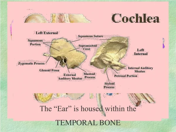
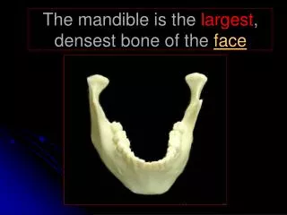
The mandible is the largest , densest bone of the face. PA Mandible. Lateral Mandible. What are the alveolar processes ?. Spongy bone on superior surface of mandible (maxillae) that presents excavation for reception of roots of teeth- Sockets.

E N D
What are the alveolar processes? Spongy bone on superior surface of mandible (maxillae) that presents excavation for reception of roots of teeth-Sockets
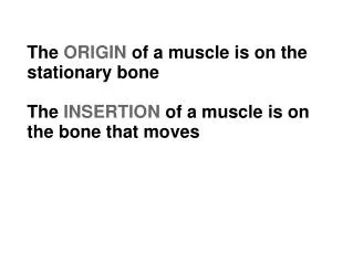
The Mandible is one of two non-paired bones of the faceWhat is the other? If you said Occipital- you are wrong! That is a cranial bone! The Vomer is the other non-paired facial bone!
Basic Routine Projections for Mandible PA Bilateral Axiolateral Obliques SMV (optional)
Notice- there is no lateral projection- why not? The two halves of mandible would be superimposed-thus obscuring information for both sides.
Mandible-PA-Ramus(General survey) Similar to a PA skull-nose and forehead against IR CR- 0 Degrees Exit at tip of nose (acanthion) 8x10 cassette, collimated (body and mentum superimposed on spine)
Evaluation Critera - PA Mandible Mandibular body and rami symmetric include entire mandible collimate Interpupillary line horizontal midsag.plane perpendicular
PA Mandible-Body • Similar to PA for Ramus, except chin and nose against IR • 0 degree CR angle
PA Mandible -Body • Ramus • Mentum • Body • Angle
A pretty decent AP mandible can be obtained by doing aC-spine odontoid R
Mandible-axiolateral oblique Both left and right oblique projections must always be performed! Blow to one side of jaw transfers force to opposite side, possibly breaking that side, not necessarily fracturing side of impact
Mandible-axiolateralobliques– 3variations • Ramus(lateral) • Body (30 degrees internally rotated) • Symphysis(45 degrees internally rotated) CR angled up 25 degrees in all projections!
Body Symphysis Ramus Mandibularaxiolateralobliques A B C
Mandibular Axiolateral oblique for Ramus Head true lateral CR angled 25 degrees up extend chin to avoid superimposition on spine
Axiolateral oblique-Ramus TMJ A CONDYLE B CORONOID C RAMUS D BODY E ANGLE F
Axiolateral oblique - MandibularBody Similar to ramus oblique, but rotate head 30 degrees to IR (mand. Body of interest will be parallel to IR) CR 25 degrees up through area of interest
Evaluation criteria -no overlap of body by opposite body no cervical spine superimposition no distortion of body Axiolateral oblique-mandibular body Coronoid • A • B • C • D • E Ramus Body Hyoid Angle
Mandibular Axiolateral oblique - symphysis No overlap of mentum no foreshortening (spine will overlap body)
Mandible-axiolateraloblique LAO LPO Shoulder super-imposition problem!
Mandible-axiolateraloblique(tip!) Why struggle to get patient into zero tilt lateral, and then angle 25 degrees up? Use tilted head to your advantage! tilt of head + CR angle 25 degrees
Mandible -SMV(optional) For visualizing mandibular body and coronoid & condyloid processes of rami
Mandible -SMV • Similar to skull, but collimated to anterior portion of cranium • IOML parallel to IR (tilt cassette forward if possible) • CR Perpendicular to IOML, midway between angles of mandible
Mandible- SMVEvaluation Criteria Symphysis A Body B Coronoid process C Distance between lateral border of skull and mand. Equal on both sides condyles anterior to pars petrosae Symphysis extendng to anterior border of face Ramus D Condyle E F Petrous ridge
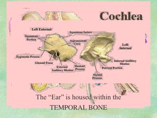
What is the only bone of the human body that does not articulate with any other bone? The Hyoid Bone!
Hyoid Bone • Attaches from the styloid processes of temporal bone to tongue • No views to demostrate
While not always necessary or useful, it is best to give the patient as much shielding as they feel they need for their safety--
REVIEW • A- Coranoid process • B- mandibular fossa • C- neck of condyle • D- Condyle • E- condylar process • F- Ramus • G- Gonion • H- Body • I-Alveolar process
1 Sphenoid bone
Sella Turcica - lateral view • Literally means “Turkish Saddle” • Superior surface of Spenoid • hypophysis = pituitary gland
Pituitary Glandsits inside Sella Turcica Once thought to be master gland- itself is regulated by hormones from hypothalamus Controls release of hormones to thyroid, gonads, adrenal cortex(growth, maturation, reproduction)
Sella Turcica series is largely obsolete! • If film readout is negative, could be diagnostic x-ray not sensitive enough to detect problem - need an MRI or CT • If positive, patient sent to CT or MRI for more detailed evaluation anyway • (CT for bone, MRI for soft tissue)
PA Axial - SellaTurcica Head and nose rest on IR ( like Skull) CR 10 degrees cephalic CR exits at Glabella Collimate to 4” X4”
Evaluation Criteria -PA Axial Sella X-ray • No rotation of cranium • Symmetric petrous ridges • Close beam restriction!
Anterior Clinoids Posterior Clinoids Dorsum Sellae Hypophysis (pituitary) Sphenoid sinus Lateral Sella Turcica A B C D E
Sella Turcica -Lateral Mid-sagittal plane is parallel with plane of IR IPL is perpendicular to IR IOML is parallel with transverse axis of film i.e.--same as Lateral skull and sinus and facial bones!
Sella Turcica -lateral cont’d Upright or semi-prone (crosstable) 8X10 cassette CR perpendicularto IR 3/4”anterior, 3/4” superior to EAM Collimate to about 4”by 4”
Evaluation Criteria - Sella Turcica Sella in center of film Sella Turcica not rotated Anterior clinoids superimposed Posterior clinoids superimposed Close beam restriction