Download
1 / 73
730 likes | 815 Views
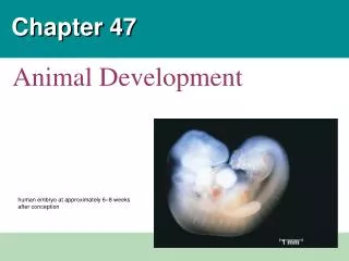
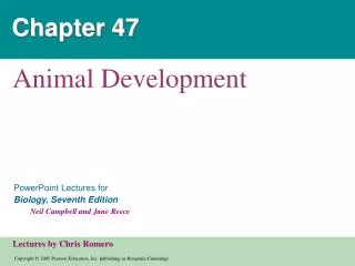
Chapter 47. Animal Development. Overview: A Body-Building Plan for Animals It is difficult to imagine That each of us began life as a single cell, a zygote. 1 mm. Figure 47.1. A human embryo at approximately 6–8 weeks after conception Shows the development of distinctive features.

E N D
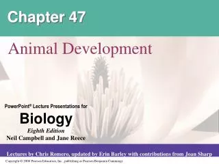
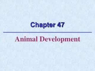
Chapter 47 Animal Development
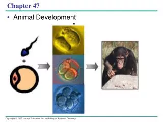
Overview: A Body-Building Plan for Animals • It is difficult to imagine • That each of us began life as a single cell, a zygote
1 mm Figure 47.1 • A human embryo at approximately 6–8 weeks after conception • Shows the development of distinctive features
The question of how a zygote becomes an animal • Has been asked for centuries • As recently as the 18th century • The prevailing theory was a notion called preformation
Figure 47.2 • Preformation is the idea that the egg or sperm contains an embryo • A preformed miniature infant, or “homunculus,” that simply becomes larger during development
An organism’s development • Is determined by the genome of the zygote and by differences that arise between early embryonic cells • Cell differentiation • Is the specialization of cells in their structure and function • Morphogenesis • Is the process by which an animal takes shape
Concept 47.1: After fertilization, embryonic development proceeds through cleavage, gastrulation, and organogenesis • Important events regulating development • Occur during fertilization and each of the three successive stages that build the animal’s body
Fertilization • The main function of fertilization • Is to bring the haploid nuclei of sperm and egg together to form a diploid zygote • Contact of the sperm with the egg’s surface • Initiates metabolic reactions within the egg that trigger the onset of embryonic development
The Acrosomal Reaction • The acrosomal reaction • Is triggered when the sperm meets the egg • Releases hydrolytic enzymes that digest material surrounding the egg
1 Acrosomal reaction. Hydrolytic enzymes released from the acrosome make a hole in the jelly coat, while growing actin filaments form the acrosomal process. This structure protrudes from the sperm head and penetrates the jelly coat, binding to receptors in the egg cell membrane that extend through the vitelline layer. 2 Contact and fusion of sperm and egg membranes. A hole is made in the vitelline layer, allowing contact and fusion of the gamete plasma membranes. The membrane becomes depolarized, resulting in the fast block to polyspermy. 3 4 Cortical reaction. Fusion of the gamete membranes triggers an increase of Ca2+ in the egg’s cytosol, causing cortical granules in the egg to fuse with the plasma membrane and discharge their contents. This leads to swelling of the perivitelline space, hardening of the vitelline layer, and clipping of sperm-binding receptors. The resulting fertilization envelope is the slow block to polyspermy. 5 Contact. The sperm cell contacts the egg’s jelly coat, triggering exocytosis from the sperm’s acrosome. Entry of sperm nucleus. Sperm plasma membrane Sperm nucleus Acrosomal process Basal body (centriole) Fertilization envelope Sperm head Fused plasma membranes Cortical granule Actin Perivitelline space Hydrolytic enzymes Acrosome Cortical granule membrane Vitelline layer Jelly coat Egg plasma membrane Sperm-binding receptors EGG CYTOPLASM Figure 47.3 • The acrosomal reaction
Gamete contact and/or fusion • Depolarizes the egg cell membrane and sets up a fast block to polyspermy
EXPERIMENT RESULTS A fluorescent dye that glows when it binds free Ca2+ was injected into unfertilized sea urchin eggs. After sea urchin sperm were added, researchers observed the eggs in a fluorescence microscope. 500 m 10 sec after fertilization 1 sec before fertilization 30 sec 20 sec Spreading wave of calcium ions Point of sperm entry CONCLUSION The release of Ca2+ from the endoplasmic reticulum into the cytosol at the site of sperm entry triggers the release of more and more Ca2+ in a wave that spreads to the other side of the cell. The entire process takes about 30 seconds. Figure 47.4 The Cortical Reaction • Fusion of egg and sperm also initiates the cortical reaction • Inducing a rise in Ca2+ that stimulates cortical granules to release their contents outside the egg
These changes cause the formation of a fertilization envelope • That functions as a slow block to polyspermy
Activation of the Egg • Another outcome of the sharp rise in Ca2+ in the egg’s cytosol • Is a substantial increase in the rates of cellular respiration and protein synthesis by the egg cell • With these rapid changes in metabolism • The egg is said to be activated
Binding of sperm to egg 1 Acrosomal reaction: plasma membrane depolarization (fast block to polyspermy) 2 3 4 6 Seconds 8 Increased intracellular calcium level 10 20 Cortical reaction begins (slow block to polyspermy) 30 40 50 Formation of fertilization envelope complete 1 2 Increased intracellular pH 3 4 5 Increased protein synthesis Minutes 10 Fusion of egg and sperm nuclei complete 20 30 Onset of DNA synthesis 40 60 Figure 47.5 First cell division 90 • In a fertilized egg of a sea urchin, a model organism • Many events occur in the activated egg
The sperm migrates through the coat of follicle cells and binds to receptor molecules in the zona pellucida of the egg. (Receptor molecules are not shown here.) 1 This binding induces the acrosomal reaction, in which the sperm releases hydrolytic enzymes into the zona pellucida. Breakdown of the zona pellucida by these enzymes allows the spermto reach the plasma membrane of the egg. Membrane proteins of the sperm bind to receptors on the egg membrane, and the two membranes fuse. 2 3 The nucleus and other components of the sperm cell enter the egg. 4 Follicle cell Enzymes released during the cortical reaction harden the zona pellucida, which now functions as a block to polyspermy. 5 Sperm basal body Zona pellucida Cortical granules Sperm nucleus Egg plasma membrane Acrosomal vesicle Figure 47.6 EGG CYTOPLASM Fertilization in Mammals • In mammalian fertilization, the cortical reaction • Modifies the zona pellucida as a slow block to polyspermy
Cleavage • Fertilization is followed by cleavage • A period of rapid cell division without growth
(d) Blastula. A single layer of cells surrounds a large blastocoel cavity. Although not visible here, the fertilization envelope is still present; the embryo will soon hatch from it and begin swimming. (b) Four-cell stage. Remnants of the mitotic spindle can be seen between the two cells that have just completed the second cleavage division. (c) Morula. After further cleavage divisions, the embryo is a multicellular ball that is still surrounded by the fertilization envelope. The blastocoel cavity has begun to form. • Cleavage partitions the cytoplasm of one large cell • Into many smaller cells called blastomeres (a) Fertilized egg. Shown here is the zygote shortly before the first cleavage division, surrounded by the fertilization envelope. The nucleus is visible in the center. Figure 47.7a–d
The eggs and zygotes of many animals, except mammals • Have a definite polarity • The polarity is defined by the distribution of yolk • With the vegetal pole having the most yolk and the animal pole having the least
The development of body axes in frogs • Is influenced by the polarity of the egg Anterior (a) Body axes. The three axes of the fully developed embryo, the tadpole, are shown above. Right Dorsal Ventral Left Posterior Animal hemisphere Animal pole Point of sperm entry 1 The polarity of the egg determines the anterior-posterior axis before fertilization. Vegetal hemisphere Vegetal pole Point of sperm entry At fertilization, the pigmented cortex slides over the underlying cytoplasm toward the point of sperm entry. This rotation (red arrow) exposes a region of lighter-colored cytoplasm, the gray crescent, which is a marker of the dorsal side. 2 Future dorsal side of tadpole Gray crescent First cleavage 3 The first cleavage division bisects the gray crescent. Once the anterior- posterior and dorsal-ventral axes are defined, so is the left-right axis. Figure 47.8a, b (b) Establishing the axes. The polarity of the egg and cortical rotation are critical in setting up the body axes.
Zygote 0.25 mm 0.25 mm 0.25 mm 2-cell stage forming Eight-cell stage (viewed from the animal pole). The large amount of yolk displaces the third cleavage toward the animal pole, forming two tiers of cells. The four cells near the animal pole (closer, in this view) are smaller than the other four cells (SEM). 4-cell stage forming 8-cell stage 0.25 mm Animal pole Animal pole Blastula (at least 128 cells). As cleavage continues, a fluid-filled cavity, the blastocoel, forms within the embryo. Because of unequal cell division due to the large amount of yolk in the vegetal hemisphere, the blastocoel is located in the animal hemisphere, as shown in the cross section. The SEM shows the outside of a blastula with about 4,000 cells, looking at the animal pole. Blasto- coel Blasto- coel Blastula (cross section) Blastula (cross section) Vegetal pole Vegetal pole • Cleavage planes usually follow a specific pattern • That is relative to the animal and vegetal poles of the zygote Figure 47.9
Fertilized egg Disk of cytoplasm Zygote. Most of the cell’s volume is yolk, with a small disk of cytoplasm located at the animal pole. 1 2 Four-cell stage. Early cell divisions are meroblastic (incomplete). The cleavage furrow extends through the cytoplasm but not through the yolk. Blastoderm. The many cleavage divisions produce the blastoderm, a mass of cells that rests on top of the yolk mass. 3 Cutaway view of the blastoderm. The cells of the blastoderm are arranged in two layers, the epiblastand hypoblast, that enclose a fluid-filled cavity, theblastocoel. Blastocoel BLASTODERM YOLK MASS Figure 47.10 Epiblast Hypoblast • Meroblastic cleavage, incomplete division of the egg • Occurs in species with yolk-rich eggs, such as reptiles and birds
Holoblastic cleavage, the complete division of the egg • Occurs in species whose eggs have little or moderate amounts of yolk, such as sea urchins and frogs
Gastrulation • The morphogenetic process called gastrulation • Rearranges the cells of a blastula into a three-layered embryo, called a gastrula, that has a primitive gut
The three layers produced by gastrulation • Are called embryonic germ layers • The ectoderm • Forms the outer layer of the gastrula • The endoderm • Lines the embryonic digestive tract • The mesoderm • Partly fills the space between the endoderm and ectoderm
Key Future ectoderm Future mesoderm 1 The blastula consists of a single layer of ciliated cells surrounding the blastocoel. Gastrulation begins with the migration of mesenchyme cells from the vegetal pole into the blastocoel. Animalpole Future endoderm Blastocoel Mesenchymecells Vegetalplate Vegetalpole The vegetal plate invaginates (buckles inward). Mesenchyme cells migrate throughout the blastocoel. 2 2 Blastocoel Filopodiapullingarchenterontip 3 Endoderm cells form the archenteron (future digestive tube). New mesenchyme cells at the tip of the tube begin to send out thin extensions (filopodia) toward the ectoderm cells of the blastocoel wall (inset, LM). Archenteron Mesenchymecells Blastopore Blastocoel Contraction of these filopodia then drags the archenteron across the blastocoel. 4 50 µm Archenteron Ectoderm Blastopore Mouth 5 Fusion of the archenteron with the blastocoel wall completes formation of the digestive tube with a mouth and an anus. The gastrula has three germ layers and is covered with cilia, which function in swimming and feeding. Mesenchyme:(mesodermforms future skeleton) Digestive tube (endoderm) Figure 47.11 Anus (from blastopore) • Gastrulation in a sea urchin • Produces an embryo with a primitive gut and three germ layers
SURFACE VIEW CROSS SECTION Animal pole 1 Gastrulation begins when a small indented crease, the dorsal lip of the blastopore, appears on one side of the blastula. The crease is formed by cells changing shape and pushing inward from the surface (invagination). Additional cells then roll inward over the dorsal lip (involution) and move into the interior, where they will form endoderm and mesoderm. Meanwhile, cells of the animal pole, the future ectoderm, change shape and begin spreading over the outer surface. Blastocoel Dorsal lip of blastopore Dorsal lip of blastopore Blastula Vegetal pole Archenteron Blastocoel shrinking The blastopore lip grows on both sides of the embryo, as more cells invaginate. When the sides of the lip meet, the blastopore forms a circle that becomes smaller as ectoderm spreads downward over the surface. Internally, continued involution expands the endoderm and mesoderm, and the archenteron begins to form; as a result, the blastocoel becomes smaller. 2 Ectoderm 3 Late in gastrulation, the endoderm-lined archenteron has completely replaced the blastocoel and the three germ layers are in place. The circular blastopore surrounds a plug of yolk-filled cells. Blastocoel remnant Mesoderm Endoderm Key Future ectoderm Future mesoderm Figure 47.12 Yolk plug Yolk plug Gastrula Future endoderm • The mechanics of gastrulation in a frog • Are more complicated than in a sea urchin
Epiblast Future ectoderm Primitive streak Migrating cells (mesoderm) Endoderm Hypoblast YOLK Figure 47.13 • Gastrulation in the chick • Is affected by the large amounts of yolk in the egg
Organogenesis • Various regions of the three embryonic germ layers • Develop into the rudiments of organs during the process of organogenesis
Neural folds LM 1 mm Neural fold Neural plate Notochord Ectoderm Mesoderm Endoderm Archenteron Neural plate formation. By the time shown here, the notochord has developed from dorsal mesoderm, and the dorsal ectoderm has thickened, forming the neural plate, in response to signals from the notochord. The neural folds are the two ridges that form the lateral edges of the neural plate. These are visible in the light micrograph of a whole embryo. (a) Figure 47.14a • Early in vertebrate organogenesis • The notochord forms from mesoderm and the neural plate forms from ectoderm
Neural fold Neural plate Neural crest Outer layer of ectoderm Neural crest Neural tube (b) Formation of the neural tube. Infolding and pinching off of the neural plate generates the neural tube. Note the neural crest cells, which will migrate and give rise to numerous structures. Figure 47.14b • The neural plate soon curves inward • Forming the neural tube
Eye Somites Tail bud SEM Neural tube 1 mm Notochord Neural crest Coelom Somite Archenteron (digestive cavity) Somites. The drawing shows an embryo after completion of the neural tube. By this time, the lateral mesoderm has begun to separate into the two tissue layers that line the coelom; the somites, formed from mesoderm, flank the notochord. In the scanning electron micrograph, a side view of a whole embryo at the tail-bud stage, part of the ectoderm has been removed, revealing the somites, which will give rise to segmental structures such as vertebrae and skeletal muscle. (c) Figure 47.14c • Mesoderm lateral to the notochord • Forms blocks called somites • Lateral to the somites • The mesoderm splits to form the coelom
Eye Forebrain Neural tube Notochord Somite Heart Coelom Archenteron Endoderm Lateral fold Blood vessels Mesoderm Ectoderm Yolk stalk Somites YOLK Yolk sac Form extraembryonic membranes Neural tube Late organogenesis. Rudiments of most major organs have already formed in this chick embryo, which is about 56 hours old and about 2–3 mm long (LM). (b) Early organogenesis. The archenteron forms when lateral folds pinch the embryo away from the yolk. The embryo remains open to the yolk, attached by the yolk stalk, about midway along its length, as shown in this cross section. The notochord, neural tube, and somites subsequently form much as they do in the frog. (a) Figure 47.15a, b • Organogenesis in the chick • Is quite similar to that in the frog
ECTODERM MESODERM ENDODERM • Epidermis of skin and itsderivatives (including sweatglands, hair follicles) • Epithelial lining of mouthand rectum • Sense receptors inepidermis • Cornea and lens of eye • Nervous system • Adrenal medulla • Tooth enamel • Epithelium or pineal andpituitary glands • Notochord • Skeletal system • Muscular system • Muscular layer of stomach, intestine, etc. • Excretory system • Circulatory and lymphaticsystems • Reproductive system(except germ cells) • Dermis of skin • Lining of body cavity • Adrenal cortex • Epithelial lining ofdigestive tract • Epithelial lining ofrespiratory system • Lining of urethra, urinarybladder, and reproductivesystem • Liver • Pancreas • Thymus • Thyroid and parathyroidglands Figure 47.16 • Many different structures • Are derived from the three embryonic germ layers during organogenesis
Developmental Adaptations of Amniotes • The embryos of birds, other reptiles, and mammals • Develop within a fluid-filled sac that is contained within a shell or the uterus • Organisms with these adaptations • Are called amniotes
Allantois. The allantois functions as a disposal sac for certain metabolic wastes produced by the embryo. The membrane of the allantois also functions with the chorion as a respiratory organ. Amnion. The amnion protects the embryo in a fluid-filled cavity that prevents dehydration and cushions mechanical shock. Embryo Amniotic cavity with amniotic fluid Albumen Yolk (nutrients) Shell Yolk sac. The yolk sac expands over the yolk, a stockpile of nutrients stored in the egg. Blood vessels in the yolk sac membrane transport nutrients from the yolk into the embryo. Other nutrients are stored in the albumen (the “egg white”). Chorion. The chorion and the membrane of the allantois exchange gases between the embryo and the surrounding air. Oxygen and carbon dioxide diffuse freely across the egg’s shell. Figure 47.17 • In these three types of organisms, the three germ layers • Also give rise to the four extraembryonic membranes that surround the developing embryo
Mammalian Development • The eggs of placental mammals • Are small and store few nutrients • Exhibit holoblastic cleavage • Show no obvious polarity
Gastrulation and organogenesis • Resemble the processes in birds and other reptiles
3 4 1 2 Endometrium (uterine lining) Inner cell mass Trophoblast Blastocoel Blastocyst reaches uterus. Expanding region of trophoblast Maternal blood vessel Epiblast Hypoblast Trophoblast Blastocyst implants. Expanding region of trophoblast Amniotic cavity Amnion Epiblast Hypoblast Chorion (from trophoblast) Extraembryonic membranes start to form and gastrulation begins. Extraembryonic mesoderm cells (from epiblast) Yolk sac (from hypoblast) Amnion Allantois Chorion Ectoderm Mesoderm Endoderm Yolk sac Gastrulation has produced a three- layered embryo with four extraembryonic membranes. Figure 47.18 Extraembryonic mesoderm • Early embryonic development in a human • Proceeds through four stages
At the completion of cleavage • The blastocyst forms • The trophoblast, the outer epithelium of the blastocyst • Initiates implantation in the uterus, and the blastocyst forms a flat disk of cells
As implantation is completed • Gastrulation begins • The extraembryonic membranes begin to form • By the end of gastrulation • The embryonic germ layers have formed
The extraembryonic membranes in mammals • Are homologous to those of birds and other reptiles and have similar functions
Concept 47.2: Morphogenesis in animals involves specific changes in cell shape, position, and adhesion • Morphogenesis is a major aspect of development in both plants and animals • But only in animals does it involve the movement of cells
The Cytoskeleton, Cell Motility, and Convergent Extension • Changes in the shape of a cell • Usually involve reorganization of the cytoskeleton
Microtubules help elongate the cells of the neural plate. Ectoderm Neural plate Microfilaments at the dorsal end of the cells may then contract,deforming the cells into wedge shapes. Cell wedging in the opposite direction causes the ectoderm to form a “hinge.” 1 2 3 4 Pinching off of the neural plate forms the neural tube. Figure 47.19 • The formation of the neural tube • Is affected by microtubules and microfilaments
The cytoskeleton also drives cell migration, or cell crawling • The active movement of cells from one place to another • In gastrulation, tissue invagination • Is caused by changes in both cell shape and cell migration
Convergence Extension Figure 47.20 • Cell crawling is also involved in convergent extension • A type of morphogenetic movement in which the cells of a tissue become narrower and longer
Roles of the Extracellular Matrix and Cell Adhesion Molecules • Fibers of the extracellular matrix • May function as tracks, directing migrating cells along particular routes
Researchers placed a strip of fibronectin on an artificial underlayer. After positioning migratory neural crest cells at one end of the strip, the researchers observed the movement of the cells in a light microscope. CONCLUSION EXPERIMENT RESULTS In this micrograph, the dashed lines indicate the edges of the fibronectin layer. Note that cells are migrating along the strip, not off of it. Direction of migration 50 µm Fibronectin helps promote cell migration, possibly by providing anchorage for the migrating cells. Figure 47.21 • Several kinds of glycoproteins, including fibronectin • Promote cell migration by providing specific molecular anchorage for moving cells
Cell adhesion molecules • Also contribute to cell migration and stable tissue structure