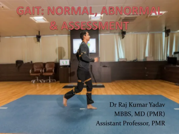
Abnormal gait
Abnormal gait . دکترامیر هوشنگ واحدی متخصص طب فیزیکی و توانبخشی قسمت 3. GAIT PATHOLOGY AND PROBABLE CAUSES 1. Foot strike to foot flat 2.Foot strike through midstance 3. Foot strike through toe off 4. Foot flat through heel off 5. Midstance through toe off 6. Swing phase.

Abnormal gait
E N D
Presentation Transcript
Abnormal gait دکترامیر هوشنگ واحدی متخصص طب فیزیکی و توانبخشی قسمت 3
GAIT PATHOLOGY AND PROBABLE CAUSES 1. Foot strike to foot flat 2.Foot strike through midstance 3. Foot strike through toe off 4. Foot flat through heel off 5. Midstance through toe off 6. Swing phase
Inadequate Dorsiflexion Control • In stance phase (Heel contact – Foot flat): Foot slap • In swing phase (mid-swing): Toe drag • Causes: Weak Tibialis Ant. Spastic plantarflexors
Flat foot strike/ Toe strike Cause: Dorsiflexor weakness Early Stance phase: • the forefoot falls to the ground without control and with a slapping sound • Foot slap gait
Toe Drag Cause: Dorsiflexor weakness Plantarflexor spasticity Swing phase: • Forefoot cannot be adequately raised and is dragged along the ground
Excessive knee extension • Loss of normal knee flexion during stance phase • Knee may go into hyperextension • Genurecurvatum: hyperextension deformity of knee • Common causes: • Quadriceps weakness (mid-stance) • Quadriceps spasticity (mid-stance) • Knee flexor weakness (end-stance) * * *
Knee Hyperextension / GenuRecurvatum • During all stance phase of gait cycle • Observe from the side view, the normal knee is completely extended only at the end of stance phase
2. Use hand to stabilize knee • Rotate the leg externally or internally • Bend trunk forward
Increased Walking Base • Normal walking base: 5-10 cm • Common causes: • Deformities • Abducted hip • Valgus knee • Instability • Cerebellar ataxia • Proprioception deficits
Walking base • During all phases of gait cycle • If distance between centre of heels is greater or smaller than normal (5-10 cm) this is classified as abnormal
Abnormal walking base • A broad walking base may be accompanied by exaggerated side-to-side displacement of the pelvis, lateral pelvic tilt, or both • An excessively narrow walking base is often caused by internal hip rotation and exaggerated knee flexion in spasticity
Wide walking base: • Contracture of hip abductors • Unsteadiness due to anxiety • Proprioceptivedeficit • General lower limb weakness • Genuvalgum • Leg length discrepancy • Pressure effects, e.g. pain or tissue damage, on the perineum
Functional Leg-Length Discrepancy • Swing leg: longer than stance leg • 4 common compensations: A. Circumduction B. Hip hiking C. Steppage D. Vaulting
Circumduction • Pesequinus • Knee / ankle ankylosis • Short contralateral • Leg Contralateral knee / hip flexion contracture
Circumduction Orothtic factor: • Knee lock • Inadequate dorsiflexion assist • Inadequate plantar flexion stop
Gait with dorsiflexor weakness: Compensatory deviation Swing phase: • Patient can compensate for this only by exaggerated hip and knee flexion • High stepping gait
Hip “hiking “ or “vaulting” • During Swing phase, when foot not contact ground and is acting like a double pendulum • Hip hiking lengthens the stride on affected side since it permits greater rotation of the pelvis in transverse plane • Helping to increase pendulum movement
Hip Hiking: • Equinus deformity or ankle dorsiflexor weakness • Skeletal shortening on sound side • It may be combined with circumduction in order to give additional anterior movement to the flexion-impaired hip joint
Gait in Cerebral Palsy Patients • Spasticity • Dynamic or fixed muscle contracture • Lever arm dysfunction • Joint contracture • Impaired balance reactions and loss of selective muscular control and equilibrium reactions, e.g. difficulty stopping if walking quickly
Common CP Knee Patterns in Gait • Crouch knee gait • Jump knee gait • Stiff knee gait • Recurvatum knee gait
Crouch Knee Characterised by: – Increased stance phase hip flexion – Persistent knee flexion >30 throughout stance – Excessive dorsiflexion throughout stance
Aetiology: – Weak ankle plantar flexors or over-lengthened TAs or from increasing height and weight – Overactive knee flexors (and/or rectus co-pasticity) – Overactive hip flexors Clinical examination – most have: – Hip flexion contractures – Knee flexion contractures – Severe hamstring tightness (popliteal angle >70)
Jump Knee Characterised by: – Increased knee flexion at initial contact correcting to near normal in mid- to late stance – Toe or flat foot strike – Increased stance phase hip flexion
Aetiology: – Overactive – hip flexors, knee flexors and/or rectus co-spasticity and/or plantar flexors Physical examination: – Usually associated with dynamic contractures – Moderate hamstring tightness (mean popliteal angle approximately 50)
Stiff Knee Gait Characterised by: – Delayed and reduced peak knee flexion in swing – Associated with compensations to aid clearance – Mainly a swing phase problem Physical examination: – Positive Duncan-Ely test – Reduced ROM in swing – Delayed and reduced peak knee flexion in swing – EMG=rectus co-spasticity in swing
Recurvatum Knee Gait Characterised by: – Toe or flat foot strike – Recurvatum >2 in stance Aetiology: – Plantar flexor over-activity/contracture – Weak dorsiflexors – Overly aggressive hamstring lengthening Physical examination: – TA tightness – Hip flexion contracture – Some hamstring tightness (popliteal angle 40)
Typical pathological gait ANKLE / FOOT • Stance phase: • No heel strike ( Flat foot contact) • Toe strike ( Forefoot contact) • Excessive plantarflexion (Vaulting) • Varus (Inversion) • Valgus (Eversion) Lack of heel rise (inadequate push off) • Swing phase: • Drop foot • Toe drag
KNEE • Stance phase: • Inadequate knee extension (flex limited/excessive) • Recurvatum (hyperextension) • Knee varus (Genuvarus) Knee valgus (Genuvalgus) • Swing phase: • Knee flexion absent • Reduced knee flexion • Increased Knee flexion
HIP • Stance or Swing phase: • Circumduction • Inadequate flexion • Inadequate extension • Exessive/ Not enough Adduction – Abduction of hip • Exessive/ Not enough External – Internal rotation of hip
PELVIS / TRUNK • Stance or Swing phase: • Anterior – Posterior tilt of pelvis • Forward – Backward trunk leaning/Lumbar lordosis • Excessive Trunk rotation • Lateral trunk bending or Trendelenberg • Hip hiking
Five gait disorders caused by cerebral lesions • frontal gait disorders • cortical-subcortical gait disorders • subcortical gait disorders • extrapyramidal gait disorders • subcorticalastatic disorders
1-Frontal gait disorders caused by • anterior cerebral artery stroke • multi-infarct state • Binswanger's disease • normal pressure hydrocephalus • bilateral frontal lobe lesions
Normal pressure hydrocephalus • CADENCE ----------Slow • STEP LENGTH-------Short • BASE ------------Slightly wide • OTHER ASSOCIATED SIGNS-------Numerous problems with handling axial body movement
Frontal lobe • CADENCE ---------- Slow • STEP LENGTH------- Slow Greatly shortened • BASE ------------ Slightly wide(protective) • OTHER ASSOCIATED SIGNS------- Difficulty starting and stopping; tendency for feet to “stick” to floor
2-Cortical-subcortical gait disorders also been termed "gait ignition failure“ "primary progressive freezing gait“ "motor blocks“ or "trepidantabasia“ Etiologies • vascular or degenerative lesions in the cerebral white and gray matter • "parkinsonian gait," - typically seen in patients with parkinsonism
3-Extrapyramidal (hyperkinetic) gait • choreic gait, dystonic gait, action myoclonus, orthostatic tremor and other hyperkinetic movement disorders • present in • Huntington's disease • idiopathic torsion dystonia • tardivedyskinesia • cerebral palsy.
4-Subcortical astatic disorders • are recognized previously as • thalamic astasia • thalamic ataxia • or subcortical disequilibrium • they are caused by thalamic or basal ganglia lesions
5-Pyramidal gait disorders • hemiparetic and spastic pattern • causes • stroke • demyelination • mass or trauma to the motor cortex or the corticospinal tracts • focal epilepsy may cause a paroxysmal gait disorder
„Cautious gait disorders„ have been labeled with many different terms • "senile gait„ • "pseudoagoraphobia," • "post-fall syndrome,„ • "space phobia," • "adaptive gait," • "astasobasophobia.„ This is the most common abnormal gait pattern in the elderly
Classification Gait Findings in Gait Disorders of Older Adults Peripheral sensory ‘‘steppage gait’ • Sensory ataxia • Vestibular ataxia • Visual ataxia Peripheral motor (‘‘Trendelenburg’’ and‘‘waddling’’ gait) • Arthritic (antalgic,joint deformity) • Myopathicandneuropathic(weakness)
Spasticity • Hemiplegia/paresis circumduction, ‘‘scissor’’ • Paraplegia/paresis Parkinsonism Cerebellar ataxia • Cautious gait • Frontal-related gait disorders, other white matter lesions