Clinical Case Conference
260 likes | 870 Views
Clinical Case Conference. David Goldberg February 16, 2011. History. Cc: Ascites and increased LAEs 64 year-old male with PMH Transfusion dependent CMML for several years S/p 1 cycle chemo (decitabine) Tongue cancer s/p chemo/xrt 2004 1 month increased abdominal distention

Clinical Case Conference
E N D
Presentation Transcript
Clinical Case Conference David Goldberg February 16, 2011
History • Cc: Ascites and increased LAEs • 64 year-old male with PMH • Transfusion dependent CMML for several years • S/p 1 cycle chemo (decitabine) • Tongue cancer s/p chemo/xrt 2004 • 1 month increased abdominal distention • Difficulty breathing • Decreased appetite • Abdominal discomfort • (+) jaundice (unclear time course) • Diarrhea x 1 month • Frequent loose BMs
History, cont’d • Reports testing “positive” for hepatitis B in 1980s • Other PMH: HTN, hyperlipidemia, PTSD • PSH: None • Meds: Amicar, Levoxyl, Flagyl, Ativan • ROS: (+) SOB • Social History: (+) 40 pack years of tobacco, “social” EtOH • Family History: No FH liver disease
Physical Exam • Vitals: BP-113/65, P-79, T-97.4 • Gen: Appeared older than stated age • HEENT: Icteric sclerae • CV: RRR, nl s1/s2, no m/r/g • Pulm: CTABl • Abd: Distended abdomen with normal bowel sounds, (+) fluid wave, (+) shifting dullness, no tenderness • Neuro: No asterixis • Skin: No palmar erythema or spider angiomata
Laboratory Values • Uric acid-7.4 • LDH-348 • CBC: WBC-4.9, Hb-10.4, Plt-28 • Chem-7: Normal, Cr-0.93 • Alk phos-716 • AST-37 • ALT-28 • Bilirubin-6.2 (3.6 direct) • Hepatitis B: SAg(-), SAb(+), CAb(+), EAb(+) • HCV Ab (-) • Ascites: 120 WBCs, 16,000 RBCs, Albumin-1.1 (serum-2.6) • INR-1.9
Questions • What is in your differential? • What would you do next in his evaluation?
Ultrasound 1. Mild hepatomegaly and splenomegaly. 2. Uncomplicated cholelithiasis. 3. Altered phasicity of the middle hepatic vein, likely secondary to diffuse infiltrative liver disease.
TJ Liver Biopsy • RA pressure: 11/0 (mean 6) mm Hg • Free hepatic vein pressure: 15/9 (mean 12) mm Hg • Wedged right hepatic vein pressure: 45/28 (mean 37) mm Hg • Corrected sinusoidal pressure: 25 mm Hg • Tryptase 582
Outline • Briefly define mastocytosis • Discuss mastocytosis of the gastrointestinal tract • Focus on hepatic complications of mastocytosis
Background • 1869: Urticaria pigmentosa decribed • 1877: Mast cells first decribed • Mastzellen: distended with cytoplasmic granules • 1949: First description of mastocytosis
Mastocytosis • Excessive mast cell accumulation • Cutaneous • Urticaria pigmentosa • Telangiectasia macularis eruptiva perstans (TMEP) • Diffuse cutaneous mastocytosis (DCM) • Solitary mastocytoma • Systemic (extracutaneous tissues) • Indolent systemic mastocytosis (ISM) • Smoldering systemic mastocytosis (SSM) • High mast cell burden • Liver or spleen enlargement but no dysfunction • Subtle signs myelodysplasia • Isolated bone marrow mastocytosis (BMM • Systemic mastocytosis with an associated hematologic non-mast cell lineage disorder (SM-AHNMD) • Associated disorder may be myeloproliferative, myelodysplastic, or lymphoproliferative. • Aggressive systemic mastocytosis (ASM) • Tissue dysfunction • Hepatic fibrosis and portal hypertension • Malabsorption • Cytopenia due to aggressive tissue infiltration • Mast cell leukemia (MCL) • Immature blasts in bone marrow or blood smear
Mastocytosis • Mast cells • Protects body from microbes and other insults • Generates inflammatory responses • Releases • histamine, heparin, leukotrienes, prostaglandins, platelet activating factor, proteases, and cytokines, including tumor necrosis factor (TNF) • Response from release • Allergic • Anaphylactic • Disease • Episodic mast cell mediator release • Chronic release • Tissue infiltration by mast cells • Associated non-mast cell clonal hematologic disease
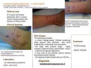
Skin findings • Darier’s sign • Urticaria and erythema within 5 minutes of scratching, rubbing, or stroking skin or skin lesions that are heavily infiltrated with mast cells Urticaria Pigmentosa: Reddish brown spots that will turn into hives when they are rubbed hard or scratched
Diagnosis • One major and one minor or three minor • Major criteria • Presence in bone marrow or extracutaneous organ of multifocal dense aggregates of greater than 15->tryptase or other special stains • Minor criteria • Atypical morphology or spindle shapes in >25 percent of the mast cells in bone marrow sections, bone marrow aspirate, or other extracutaneous tissues • Mutational analysis of KIT showing a codon 816 mutation (eg, Asp816Val) in bone marrow, blood, or extracutaneous organs • Bone marrow or other extracutaneous mast cells expressing the surface markers CD2, CD25, or both • Serum tryptase levels (when the patient is in a baseline state) >20 ng/mL.
GI tract and SM • 16 consecutive patients with systemic mastocytosis • 9 w/ DU or duodenitis • Gastric acid hypersecretion in 6 • Mean BAO 20.7 mEq/h • Increase plasma histamine • Correlated with BAO • Decreased gastrin • Abdominal pain or diarrhea • 80% patients • PUD: 15-44% Cherner et al. Gastroenterology 1988
GI tract and SM • 14-85% cases • 16 series • Since 1985: 60-80% • GI symptoms • Abdominal pain (51%) • Diarrhea (43%) • Nausea/vomiting (28%) • Secondary to mast cell mediators Lee et al. World J Gastro 2008
GI tract and SM • Abdominal pain • Epigastric: Ulcer disease and acid hypersecretion • Lower abdominal: Cramping due to motility • Diarrhea • Gastric acid hypersecretion • Volume of gastric acid • Mucosal injury/ulceration->secretory diarrhea • Inhibits pancreatic enzymes • Steatorrhea->lipase • Altered bowel motility • Malabsorption • Mucosal injury • Edema Lee et al. World J Gastro 2008
Flushing • Carcinoid • Patchy • Violaceous • Lasts few minutes • Postprandial • Not associated with hemodynamic changes • Systemic mastocytosis • Bright red • Pruritic • Burning • Other symptoms of mast cell degranulation Lee et al. World J Gastro 2008
Liver disease and SM • Hepatomegaly • 41-72% • More commonly seen in more advanced/aggresisive disease • Cirrhosis • 4% patients • LAE abnormalities • Alk phos more common • Correlates with severity of disease, degree of fibrosis, and mast cell load • Portal venopathy or VOD Lee et al. World J Gastro 2008
Portal Hypertension • Portal hypertension • Intrahepatic venous obstruction • Fibrosis in portal areas or hepatic sinusoids • Pre- and post-sinusoidal blockage secondary to mast cell infiltration and/or fibrosis • Increased splenic vein blood flow • AV shunts in spleen->vasodilators • Hepatic venous outflow obstruction • Occurs in 10-40% of patients • Ascites common Jensen R et al. Hematology/Oncology Clinics of North America 2000
Liver disease and SM • 182 patients with generalized mastocytosis • 52 own cases 130 from literature • 131 (72%) with hepatomegaly • 25 (14%) with periportal fibrosis • 7 (4%) with cirrhosis • 77 (42%) with confirmed mast cell infiltration histologically • Predominantly in portal tracts • Loosely scattered throughout sinusoids Horny et al. Cancer 1989.
Liver disease and SM • Portal hypertension • 4 cases SM, 3 with portal HTN and ascites1 • No cirrhosis on liver biopsy • Lab findings c/w autoimmune cholangitis • Non-cirrhotic portal HTN->erroneous diagnosis of cirrhosis • 49 F p/w 6 weeks ascites • Liver biopsy w/ periportal fibrosis • Sinusoidal infiltrate suggestive of myeloid leukaemia • Centrilobular cell loss and congestion c/w hepatic venous outflow obstruction • Initially diagnosed with CMML • Toluidine blue stain showed mast cells in portal tracts • Intrahepatic cholestasis • 35 F w/ mast cells in portal tracts and centrizonal cholestasis3 • Nl ERCP • Cholestatic labs resolved with chemo 1-Kyriakou et al. Am J Gastro 1998; 2-Naryanan Post Grad Med Journal 1989; 3-Safyan et al. Am J Gastro 1997
Treatment • H2 blockers (gastric acid hypersecretion) • Dyspeptic pain • Diarrhea • Malabsorption • Higher dose • Cromolyn sodium • Mast cell membrane stabilizer • Diarrhea • Abdominal pain • Cramps
Conclusions • Mastocytosis is a rare disorder resulting from excessive mast cell accumulation • GI tract can be involved, leading to PUD and diarrhea • Liver commonly involved in SM, and can result in portal hypertension, cirrhosis, and associated complications