Download
1 / 30
300 likes | 391 Views
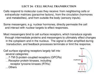
MPHY616. Signal Transduction and Cell Cycle Paul Shapiro, Room 222 Pharmacy Hall pshapiro@rx.umaryland.edu Office: 6-8522 Lab (Rm 269): 6-2980. Objectives: A. Overview cell cycle. B. Define major mitotic structural changes. C. Define major G2/M-phase signaling events. Somatic Cell Cycle:.

E N D
MPHY616 Signal Transduction and Cell Cycle Paul Shapiro, Room 222 Pharmacy Hall pshapiro@rx.umaryland.edu Office: 6-8522 Lab (Rm 269): 6-2980 Objectives: A. Overview cell cycle. B. Define major mitotic structural changes. C. Define major G2/M-phase signaling events.
Somatic Cell Cycle: G0 R R = restriction point
Mitosis: Prophase Metaphase Anaphase Telophase Entry: *Chromosome condensation *Spindle pole formation *Nuclear envelope breakdown *Other structural changes Exit: *Chromosome congression *Microtubule attachment *Chromosome segregation *Nuclear envelope reassembly *Other structural reassembly
Regulation of CDK activity. • Cyclin synthesis and binding • CDK phosphorylation • CDK dephosphorylation
Cdc25A Cell Cycle: G1-Phase Cdk4/6 CyclinD (+) • Cdk 4/6 activated through interaction with cyclin D and dephosphorylation. • Rb phosphorylation relieves Txn repression. • Expression of genes required for DNA synthesis. Cdk2 (+) CyclinE Rb Inactive Active pRb HDAC Rb X Transcription E2F E2F DP-1 DP-1 OFF ON
Cell Cycle: G1-Phase • G1 progression: Dependent on mitogen availability. • Increased expression of cyclin D, immediate early genes • (Fos, Myc). Dependent on ERK MAP kinases (TCF/Elk-1). • Activation of protein translation: Indirect phosphorylation • of eIF4E by ERK. Enhances mRNA binding and protein • expression. • G1 inhibition: G1/S checkpoint • (No mitogens, senescence, contact inhibition, DNA damage). • A. Increased expression of Cdk inhibitors (CdkI) • -p15/16 INK4 target Cdk4/6 • -p21CIP / p27KIP target Cdk2 • *ERK and p53 (DNA damage) up-regulate p21CIP • expression. [p53 regulation by phosphorylation] • B. Increased cyclin D degradation: GSK3b phosphorylation • Decreased Cdc25A expression: TGF-b mediated.
G1 DNA damage checkpoint. • DNA damage / recognition. • Phosphorylation • Stabilization of p53 • Expression of p21 • Inactivation of CDK
Cell Cycle: S–Phase / DNA synthesis • 1. Accurately replicate each chromosome. • (avg. 40mm DNA / Chromosome) • *Replication initiated at multiple • sites on chromosome. • Reassemble into proper structure. • *Nucleosomes / Scaffolds. • Dependency of M-phase on S-phase. • *Unreplicated or damaged DNA. • Dependency of S-phase on M-phase. • *Prevent changes in cell ploidy. • Endoreduplication / endomitosis: • *Successive S-phases without M-phase.
Cell Cycle: G2-Phase progression Goal: Prevent transmission of DNA replication errors or DNA damage and prepare for mitosis. Myt1 and Wee1 Cdc25B/C (+) (-) Cdc2+ CyclinB Tyr15 (-) (+) G2-Phase Mitosis
Cell Cycle: G2-Phase (DNA damage checkpoint) *DNA damage *Sensors *Effectors and co-effectors DNA-PK ATM/ATR Chk1/2 p53 p21CIP, GADD45 Cdc25B/C (-) Cdc2/CyclinB *G2 progression blocked
Stages of mitosis: Start 4 meters of DNA in 46 chromosomes per G2 cell.
The Microtubule Organizing Center: A. Animal cells: centrosome B. Yeast: spindle pole body. Major Components: 1) Centriole pair surrounded by pericentriolar material. 2) g-tubulin - acts as tether for a and b tubulin and microtubule nucleation (+ and - ends). 3) Motor proteins: Dynein and kinesin. (+ accessory proteins)
Centrosomes and Centrioles: 1) Duplication at G1/S boundary is distinct from maturation during G2/M transitions. A) Control of duplication: Protein kinases Mps1p (S. cerevisiae), Cyclin E and A/cdk2. B) Centrosome maturation: growth in size during G2/M. -Recruitment of cytoplasmic proteins. -Nucleation of microtubules. Centrosome proteins: Maybe ~50, including signaling proteins, chaperones, motor proteins, structural proteins.
Centrosome maturation: role of phosphorylation. A. Regulation by protein kinases: Polo-like kinases (Plk). -Human homologue to Drosophila polo gene product. -Plk active during M-phase and may regulate entry and exit. B. Plk regulation of G2/M transitions: Microinjection studies showed profound effects on centrosome maturation and bipolar spindle formation. *Differences between normal diploid cells and tumor cells suggested the presence of a centrosome checkpoint. C. Plk is regulated by upstream kinases: D. Potential target proteins: -Centrosome? -Cdc25 -Anaphase promoting complex (APC) (3 proteins: Cdc16,27, and Tsg24)
Spindle Assembly Checkpoint: (also called mitotic or kinetochore-attachment checkpoint) Function: Inhibit progression into anaphase in order to prevent chromosome segregation errors. Questions: 1) What senses proper MT attachment or tension? 2) How does one unattached chromosome inhibit anaphase in the attached chromosomes? Figure from: W. Wells (1996) Trends in Cell biology, Vol 6: p228.
Spindle checkpoint proteins: MAD (mitotic arrest-deficient) BUB (budding uninhibited by benzimidazole) MAD1/2/3 - All interact with anaphase promoting complex proteins (at least in 2-hybrid screens) and are thought to inhibit. MAD1-phosphoprotein (by Mps1?) (pre-meta kinetochores). MAD2-cointeracts with MAD1 (pre-meta kinetochores). MAD3-unknown function. BUB1/2/3 - BUB1 - S/T kinase (pre-meta kinetochores). BUB2 - unknown function. BUB3 - associates with BUB1, BUB1 substrate (pre-meta kinetochores).
Mad2 at unattached kinteochores Red = unattached White = just attached
Nuclear Envelope Breakdown (NEB) during mitosis: • Nuclear envelope versus nuclear membrane: • Envelope consists of: • Nuclear pore complex • Outer membrane • Inner membrane • Lamina: • -(A/C and B-type lamins) • -lamina-associated • polypeptides (LAPs) • -lamin B receptor (LBR)
B. Rapid NEB at the end of prophase. C. NEB regulated by phosphorylation of lamins and other lamina proteins: Not simply due to increases in cdc2/cyclin B activity. Other lamin kinases: PKC, Arg-Ser (RS) kinase, PKA D. Reassembly of nuclear envelope following dephosphorylation: Protein Phosphatase 1 (PP1) targeting of lamin B involved.
NEB: Regulation may involve functional nuclear pores and lamina. A. Lamin B and p56 (inner membrane protein and putative lamin B receptor) interactions weakened by hyperphosphorylation. B. Intact nuclear pore complexes may be required for hyperphosphorylation. C. Similarly, nuclei disassembly does not occur without lamina. D. Pores likely provide access to lamin kinases. (+) mitotic extracts From Collas (1998) J. Cell Science, Vol.111:1293.
Organelle disassembly during mitosis: Golgi fragmentation Warren (Yale): Cdc2 mediated GM130 phosphorylation and p115 interactions. Malhotra (UCSD): Raf-1 and MEK1 activity. Targets?
Mitotic Exit: Anaphase Promoting Complex (APC) / Cyclosome. 1) Composed of at least 8 subunits in vertebrates. 2) Catalyzes the transfer (ligation) of ubiquitins to a N-terminus 9 amino acid destruction box (D-box) sequence on cyclins and other proteins. (RXALGXIXN) E1: ubiquitin-activating enzyme. E2: ubiquitin carrier. E3: ubiquitin protein ligase. 3) Ubiquitinated proteins targeted for degradation. 4) APC activity regulated, in part, by phosphorylation. (Kinases involved and mechanisms not well defined) 5) Protein-protein interactions involved in APC activation. (ATP-independent)
Regulators of APC activity during metaphase: WD40 repeat proteins - Humans; hCDC20 (p55CDC) and hCDH1 S. Cer. (Cdc20p, Cdh1p/Hct1p) D. mel. (fizzy, fzy and fizzy-related, fzr) CDC20 and CDH1 bind APC and activate cyclin ubiquitination activity. CDC20 regulates APC in mitosis and confers a strict D-box dependence on APC, CDH1 is not strictly dependent on D-box. CDC20 protein levels increase in G2/M, whereas, CDH1 levels are relatively constant throughout the cell cycle. CDH1 may facilitate G1 degradation of proteins lacking D-box (eg. Plk).
Model for cell cycle regulation of APC: From Fang et al., Mol. Cell (1998) Vol.2:163 (M. Kirschner-Harvard) Degradation Cyclin B/Cdc2 Other kinases? Mitotic Checkpoint CDC20 CDH1 CDC20 and P CDC20/CDH1 APC-P CDH1 APC APC APC-P S/G2 Metaphase Early Anaphase Late Mitosis/G1