Download
1 / 23
250 likes | 786 Views
Valvular Heart disease HVD. By Dr. Ashraf Abdelfatah Deyab. VHD- Objectives. By the end of this session, the student should be able to: Define and classify valvular heart disease. Enlist the causes of acquired heart valve diseases.

E N D
Valvular Heart disease HVD By Dr. AshrafAbdelfatahDeyab
VHD- Objectives • By the end of this session, the student should be able to: • Define and classify valvular heart disease. • Enlist the causes of acquired heart valve diseases. • Identify the clinical consequences of valve dysfunction and complications. • Describe different morphological features of valve dysfunction.
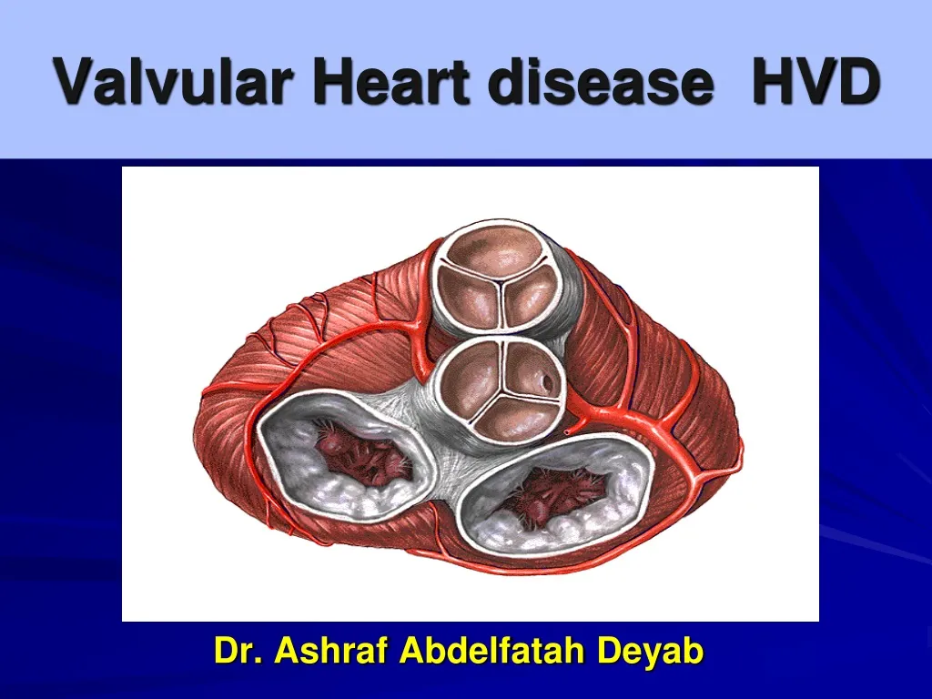
Valvular Heart Disease(HVD) • Function of normal Valves – • Unidirectional blood flow, one-way flow of blood from the atria to the ventricles to the arteries. • Name of heart valves – • 1.Two atrioventricular valves: • Mitral valve: Left heart - “Bicuspid valve” . • Tricuspid valve:Right heart -“tricuspid” • 2. Two semilunar valves: • Aortic valve: Left heart . • Pulmonary valve: Right heart. • Valve competency depends on – • 1. Annulus, 2. Leaflets, 3. Cords, 4. Papillary muscles, 5.Ventricular wall layers
The mitral valve The aortic valve Bicuspid valve-thin delicate& leaflets Three thin and delicate cusps
Define HVD, and explain why its draw the clinical attention? • HVD is groups of critical clinical conditions involve heart valves, leading to different pattern of dysfunction. • HVD come to clinical attention – because impose: • Hemodynamicinstability. • Increase susceptibility to infection (infective endocarditis). • Why hemodynamic burden precipitated?
Abnormal Valve Function • 1. Valve Stenosis • Obstruction to valve flow. • 2. Valve Regurgitation, Insufficiency, Incompetence • Inadequate valve closure--- back leakage. • 3. A single valve can be both stenotic and regurgitant; but both lesions cannot be severe!! • 4. Combinations of valve lesions can coexist • Single disease process • Different disease processes • One valve lesion may cause another
Abnormal valve function • Definition of Valvular stenosis ? • Stenosis is the failure of a valve to open completely, which obstructing forward flow. • Etiology • Almost caused by chronic primary cuspal abnormalities- (1)Calcification or (2)Valve scarring. • Stenosis of the mitral valve is a common complication of rheumatic fever. • Definition of Valvular Regurgitation ? • Insufficiency results from failure of a valve to close completely, thereby allowing reversed flow. • Etiology • (1) Intrinsic disease of the valve cusps= destruction. • (2) Distortion of supporting structure (papillary M, cords,etc.)
Classification • Based on etiology can be classified into: • 1. Congenital heart disease • 2. Acquired heart disease.
Heart Valvular Disease- Etiology 1.Congenital heart valve disease - e.g. Septal defect, Atresia, mal-position. to be discussed in separate session. _______________________________________________ 2. Acquired heart valve disease :- (most frequent) • Endocarditis– (MR & AR) most common is mitral valve. • Post-inflammatory healed scar (Rheumatic heart disease) MS+MR & AS+AR • Senile calcific aortic stenosis-AS • Myxomatous - Mitral valve Prolapse- MR • Abnormalities of Leaflets and Commissures • Abnormalities of Tensor Apparatus. • Abnormalities of Left Ventricular Cavity and/or Annulus-
Valvular Heart Disease- Clinical consequences The clinical consequences depend on : • Type of valve involve. • Degree of impairment. • How fast it develops. (Acute form and chronic form) • Rate of compensatory mechanism. Clinical Outcomes: • 1) Stenosis leads to pressure overload of the heart. • 2) Insufficiency leads to volume overload of the heart.
VALVULAR STENOSIS Pressure in upstream chamber IS HIGHER than Pressure in downstream chamber during time of flow (when valve is normally open). Hemodynamic abnormality = "PRESSURE GRADIENT" Upstream Down stream High pressure low pressure
VALVULAR REGURGITATION Retrograde flow of blood "upstream" during time when valve is normally closed. Hemodynamic abnormality = "VOLUME OVERLOAD" Upstream Down stream Volume overload
Assessment for Valve Dysfunction • Murmurs • General malaise • Dyspnea on exertion • Dizziness • Chest pain or discomfort • Prior history of rheumatic heart disease • Orthopnea • Dyspnea, rales • Pink-tinged sputum Complications: • Hemodynamic instability • Heart failure • Angina • Syncope • Death Diagnosis: • ECG • Chest x-ray • Cardiac cath • Echocardiogram
Heart Valvular Disease- Clinical Outcomes • Example: • (1) Mitral stenosis: (comments type) • Complication of Rheumatic heart disease fibrotic\scarring • Chronic - Well tolerated over years. Mitral stenosis “fish mouth”show diffuse fibrous thickening &distortion, commissural fusion
Calcific aortic disease • Most common acquired aortic stenosis in elderly. • Consequence of age-associated “wear and tear” degeneration , fibrosis and calcification. • Occasions: (1) Normal valves. (2) Congenitally bicuspid valves • Pathological processes for calcification (1) Disorder of elderly (2) Unknown. • The major clinical features of Stenosis : • (1) Left ventricular hypertrophy and (CHF) failure... • (2) Angina. • (3) Syncope (abrupt episodes of faintness) (hypoperfusion)
Calcified aortic valve of old age Macroscopic • Heaped-up protruded • calcified masses. • 2) the cusps become fibrosed • and thickened. • 3) The free edges of the cusps • are not involved. Microscopic: • large nodular calcific deposits.
MITRAL VALVE PROLAPSE (MPV) • Definition: • Mitral valve leaflets (one or both) are “floppy” and Prolapse, or balloon back, into the left atrium during systole. • The histologic change in the tissue is called myxomatous degeneration. • MVP-Uncommon, affects approximately 3% of adults in USA. • Women 7times more frequently > Male • Pathogenesis of MVP: • (1)Unknown, • (2) MVP is associated with heritable disorders of CT diseases Marfan syndrome (fibrillin-1 mutation), where there is intrinsic defect of CT either in its Synthesis or Remodeling.
MPV-MORPHOLOGY • Macroscopic appearance • The Leaflets: Enlarged, redundant, thick, rubbery, Ballooning . • The Tendinous : cords may be elongated, thinned, or even ruptured. • The annulus: may be dilated. • The tricuspid, aortic, or pulmonary valves may also be affected. Left ventricle demonstrating prolapse of the posterior mitral leaflet into the left atrium
Mitral valve Pronounced hooding of MV with thrombotic plaques
Microscopy: * Thinningfibrosa layer of the valve. * Markedexpansion of the spongiosa layer with deposition of mucoid (myxomatous) material.
1. Stenosis is the failure of a valve to close completely. (T) OR (F) • 2. Insufficiency is the failure of a valve to close completely. (T) OR (F)


