Introduction to Anatomical Planes, Sections, and Epithelial Tissue Classification
This lab manual covers essential anatomical terminology, including sagittal, frontal, and transverse planes, along with directional terms vital for understanding human anatomy. It also explores body regions and the classification of tissues, with specific emphasis on epithelial types like stratified squamous and simple columnar. Students will sketch various tissues, noting their characteristics and locations, due by 6/25. Understanding these fundamental concepts is key for further study in histology and tissue functions related to protection, absorption, and secretion.


Introduction to Anatomical Planes, Sections, and Epithelial Tissue Classification
E N D
Presentation Transcript
Planes and Sections see xv in lab manual • Sagital • Mid-sagital • Para-sagital • Frontal • Transverse
Directional Terms – See xiv in lab manual • Right - Left • Medial – Lateral • Distal – Proximal • Dorsal – Ventral = Anterior – Posterior • Superior – Inferior = Cranial – Caudal • Superficial - Deep
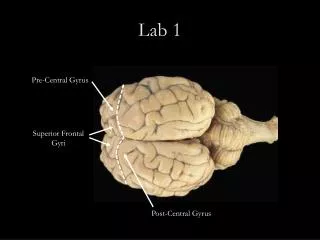
Axial Cephalic Cervical Thoracic axillary Abdominal Lumbar Pelvic inguinal Appendicular Acromial Brachial Cubital Antebrachial Carpal Palmar gluteal Femoral Popliteal Crural Sural Tarsal Body Regions
Tissues • Epithelial • Connective • Muscle • Nervous
HW - 1 • Sketches of specific tissues • No more than 4 per page • Single sided • List major characteristics and where typically found • Due 6/25 beginning of lab – prior to exam • Diminished credit if turned in late
Epithelial p 56+ in text • Common characteristics • Types • Keratinized and nonkeratinized stratified squamous • Simple squamous • Simple columnar • Simple Cuboidal • Pseudostratified ciliated columnar
Epithelial Tissue Functions: protection, absorption, secretion Cover all virtually all body surfaces, in and out Lack vascular supply Classified based on shape and stacking of cells Generally supported on a basement membrane (& connective tissue)
Classification of epithelial tissues 1) Cell shape (at free surface) a) squamous - flat cells b) cuboidal - cube shaped c) columnar - column shaped Reminders: -histology = thin section of a 3D object -there are MANY tissues on each slide
Classification of epithelial tissues 2) Cell layers/arrangement a) simple - a single layer - relatively thin/fragile - found lining internal structures - simple squamous epithelium pictured here b) stratified -multiple layers -thicker -may be in areas w/ stress -stratified squamous epithelium w/out keratin pictured here
Name this epithelial tissue: Cell arrangement/layers…….Shape of cells at surface
Pseudostratified Columnar (Fig 3.6.b): -Lining much of the respiratory tract -often have cilia & goblet cells -looks layered BUT there is really only 1 layer of cells with nuclei at different heights
Name this epithelial tissue: Cell arrangement/layers…….Shape of cells at surface
Name this epithelial tissue: Figure 3.6b
Connective p 64+ in text • Common characteristics • Types • Dense • Cartilage • Bone • areolar • Adipose • Blood
Connective Tissue Most abundant tissue in body Not on free surfaces Cells separated in a matrix (ground substance + fibers) Good nerve & blood supply (except cartilage & tendons- dense connective) Classified based on matrix components, arrangement and type of protein fibers