Download
1 / 62
640 likes | 808 Views
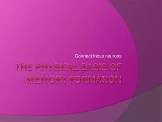
Connect those neurons. The physical basis of memory formation. NEURAL BASIS OF MEMORY. Memories are stored throughout the brain and linked together through neural tracts or pathways, often described as “memory circuits” consisting of interconnected neurons.

E N D
Connect those neurons The physical basis of memory formation
NEURAL BASIS OF MEMORY • Memories are stored throughout the brain and linked together through neural tracts or pathways, often described as “memory circuits” consisting of interconnected neurons. • Not all areas of the brain are equally involved in memory, certain brain areas are more or less involved in different memory processes. • E.g. Temporal lobe – important role in declarative explicit memories (“knowing that”) but are less important for procedural implicit memories.
The Role of the neuron in memory formation • New memories (either short or long term) are NOT stored in individual synapses but in the pattern of thousands of new interrelated connections • Looking for memories in a single nerve cell or synapse is a dead end • We know that there is a molecular basis to memory formation, what we do not know is exactly how thousands of these new connections hold our memories.
ERIC KANDEL • Many studies have been done on how brain neurons change when a new memory is formed – the most influential conducted by Austrian born US psychiatrist and neuroscientist Eric Richard Kandel over the past 50 years. • Kandel has identified changes in the structure and functioning of neurons in the brain when forming the memory of a newly learned experience. • Kandel was awarded Nobel Prize in Physiology or Medicine in 2000.
The role of the neuron in memory formation • Eric Kandel studied the sea slug AplysiaCalifornica (a seaweed munching seaslug found off the coast of California) Why a seaslug? • Very simple nervous system – far less complex than human and other mammals • 20,000 neurons – compared to trillions of neurons in human brain and other mammals such as monkeys and cats. • Largest neurons in the world that can be seen with the naked eye • Neurons can be observed for days without any difficulty. • Neurons can be easily stimulated to observe neuronal changes or removed one at a time for biochemical analysis.
Kandel’s experiments • Stimulated the siphon (gill in the tail of slug that squirts water to move slug away from danger) with a very thin electrode or “probe”. • STM – would withdraw gill more and more quickly • Forgetting – an hour later the withdrawal was again slow, progressively faster with continued stimulation • Habituation – eventually the slug stopped responding to the stimulation as it caused no damage, it had ‘learned’ that the shock was harmless
Kandel’s experiments • Each day the slug would habituate more quickly than the day before • This suggest some kind of LTM lasting days or even weeks • By studying the neurons involved in this process he identified the changes that allowed the learning to take place • The neurons were physically changing! • These changes are called collectively Long Term Potentiation
Neuronal changes 1.The way the neurons function. There is an increase in the amount of neurotransmitter produced and released by the neurons. (i.e. The specific chemical substance used by neurons to communicate) 2. The structure of the neurons. The number of axons (send messages) and dendrites (receive messages) increases as they become “bushier” through the growth of smaller offshoots (called dendritic spines) thus strengthening the connections between neurons. 3. Involves the synapse (the small space between the axon of one neuron and the dendrite of another). When a memory is formed, new synaptic connections form (called “synaptic growth”) and this further strengthens the connections between the neurons, making it easier for them to transmit to each other the next time, thus enabling communication to occur more easily.
Neuronal changes • The more neurons in a circuit are activated through use, the “easier” it becomes for information to travel through the circuit. • All of these changes are called “LONG TERM POTENTIATION” (LTP).
Long Term Potentiation • Long term potentiationrefers to the longlasting strengthening of synaptic connections of neurons resulting in the enhanced functioning of the neurons. • Neural basis for memory formation • Synapse strength can increase in 3 ways • Release extra neurotransmitter • Increase number of receptor sites • Growth of new synapses
SUMMARY OF KANDEL’S FINDINGS • Any experience that results in memory produces physical changes in the brain at the neuronal level, strengthening connections between neurons involved in the process, thus making communication easier next time. • The neuronal changes create and strengthen a memory circuit of information that has been learned. • With STM storage, there is only an increase in the release of neurotransmitter. • With LTM storage, all structural and functional changes occur. Memory circuit is activated each time a memory is recalled. • Difficult to generalise sea slug research findings to humans. • Difficult to study long term potentiation in humans due to complexity of our brain and its neural circuits. • There is other research that shows long term potentiation is associated with memory formation in fish, chicks, mice, rats, cats, rabbits and squirrels.
Long Term Potentiation– Explicit STM New Receptor Formation Stronger neural impulse in post synaptic neuron
Long Term Potentiation Short Term Memory New Receptor Formation Late LTP Long Term Memory New Synapse Formation
ROLE OF HIPPOCAMPUS • Just above each ear and about 4 cm straight into the brain is the hippocampus. • It is a tubular, curved, somewhat sea horse shaped structure. • In humans, it is about 3.5 cm long and we have two of them (one in each lower region of the temporal lobe in each hemisphere).
The Medial Temporal Lobe • The medial temporal lobe is the inner surface area towards the middle (“medial”) of the temporal lobe that includes the hippocampus, the amygdala, and other cortical tissue.
The Hippocampus & Medial Temporal Lobe – damage and memory • Henry Molaison patient in 1957 study (H.M.) then participated in hundreds of studies until he died in 2008 at age 82. • Severe epilepsy since age 10 so at age 27 he agreed to brain surgery. • Radical surgery removes hippocampus and parts of the medial temporal lobe because H.M.’s seizures were so severe and precise origin could not be determined. • Removed about 5 cm of tissue from each lobe. • Surgery was a success in reducing frequency and severity of seizures and could also be controlled with medication. • Personality basically unchanged. • Many cognitive functions remained unaffected. • Left with permanent anterograde amnesia (Cant form new LTM’s) • Other mental abilities and STM fine
H.M. case • Could conduct a conversation as normal as long as not distracted. • Had a good vocabulary, normal language skills, slightly above average intelligence. • Left with serious memory problems. • H.M. could remember events that occurred well before his surgery but could not remember things he experienced after his surgery. E.g. Could remember childhood events but not what he had for breakfast. • He had become incapable of forming new long term episodic (personal events) or semantic (general knowledge) memories. • He had to be reintroduced to his doctors every time they visited, including his neuropsychologist who tested him regularly for over 50 years.
H.M. case • He forgot the news almost as soon as he had seen or heard something. Had no idea of the time of day unless he had just looked at the clock. Each time he was told his uncle had died he reacted with surprise and became just as upset as when he was first told. • STM was normal. He could still learn new motor skills or do crossword puzzles. As long as he paid attention to a task and thought about it or actively rehearsed it he could retain information in STM. But when distracted, turning his attention to something else, he immediately forgot about it – the information vanished without a trace and could not be recalled thereafter.
The Hippocampus & Medial Temporal Lobe – damage and memory • Evidence that the hippocampus and temporal lobe have a role in memory formation, but not storage or retrieval • Hippocampal area of temporal lobe has an important role in formation or encoding of new declarative explicit memories (semantic and episodic) but not in formation or retrieval of implicit procedural memories. • Provides evidence that LTM involves distinctive or relatively “independent” storage and retrieval processes. • Evidence that LTM is most definitely a distinct sub system of memory (STM fine) • Evidence that the hippocampal area of the temporal lobe is not involved in short term storage.
Current research • No longer perform surgery to remove both medial temporal lobes to treat epilepsy. • Case studies of patients with injury or disease to hippocampal area in both lobes indicate they experience the same type of amnesia (anterograde amnesia) and difficulties forming new long-term explicit memories. • Experimental research using neuroimaging techniques has also provided evidence of these roles of the hippocampal area.
Why anterograde amnesia? • Psychologists have proposed that the hippocampus acts as a kind of “memory formation area” where the brain temporarily holds and processes components of the information to be remembered. E.g. Rock concert – venue, friends you were with, visual images, band members, songs. • The different components need to be integrated or linked together in the hippocampus to form a single episodic memory. • When the memory is formed the different components will then be gradually transferred to cortical areas in the brain specialised in the long term storage of that specific type of information. (This may also be why H.M. Could retrieve old memories – the memories were located elsewhere in the cortex and had already been formed with well-established circuits linking the components and so were not dependent on the hippocampal area).
Deep within the temporal lobe- the amygdala • Mediation of fear – sympathetic arousal • Seizures involving the amygdala involve intense fear • Damage leaves a person unable to learn a fear response through classical conditioning • Involved in remembering the emotional significance of an event • Can effect the consolidation of memory – stimulation better recall, retardation poorer recall
Consolidation theory • Some research evidence suggests that new information transferred from STM to LTM requires a period of time for consolidation or strengthening in order for it to be encoded and permanently stored. • Consolidation theory proposes that structural, or “physical” changes to the neurons in the brain occur when something new is being learned and immediately following learning.
Consolidation Theory • Physiological neuronal changes (LTP and Axon growth) in brain cells occur when something is being learned and during a period of time immediately after the learning process has been completed • If memory is disrupted during the period of consolidation then information may not be embedded in LTM and will therefore be lost. • If there is no disruption, then information becomes a permanent part of LTM, at least until retrieved. • Consolidation is a gradual process and material being remembered is vulnerable to disruption for at least 30 minutes, although consolidation of memories can proceed for as long as several years in people and for weeks in animals such as mice, rats and rabbits (Zimbardo, 1992) • This process takes about an hour
Consolidation Theory • Hippocampus and Medial Temporal Lobe involved – in H.M.’s case when these were removed he was unable to consolidate new memories due to lack of structures that co-ordinate the strengthening of the neurons and neural connections involved in the new memories. • It is aided by REM sleep • Think of wet concrete needing to “set”
Evidence for Consolidation theory • Studies of people who have experienced brain trauma resulting in memory loss – e.g. Knocked unconscious after accident , after acquiring brain disease (such as encephalitis) or after receiving electroconvulsive shock theory (ECT) as treatment for severe depression. • The above cases see people frequently unable to report any memory of events immediately before the accident or treatment, even for up to 30 minutes before the brain trauma. • Neural changes are not allowed to occur or are disrupted by injury
Consolidation theory research • Other evidence comes from research with animals. Rats given ECT at various time intervals after learning to run a maze would remember the task they had learned differently. (Hudspeth, McCaugh & Thompson, 1964) • Group A ECT immediately after learning task • Group B ECT 20 secs after learning • Group C ECT 30 minutes later • Group D ECT 60 minutes later Results showed consolidation occurred within about 60 minutes after learning. None of rats in Group A remembered the task Partial retention for rats in Groups B and C with C being better retention than Group B All rats in Group D remembered the task completely.
Reconsolidation • After a memory is activated and retrieved from LTM it needs to be consolidated again in order to be stored back in LTM. This process is known as reconsolidation. • (Gazzaniga and Heatherton, 2006) Evidence from rats injected with drugs when recalling previously consolidated LTM’s became unable to recall what they had previously demonstrated knowledge of. This suggests that memories for past events can be affected by new circumstances once they are retrieved, so that newly reconsolidated memories may differ from their original versions. e.g. Borrowed book from library with some pages torn out – some information is no longer retrievable so book placed back on shelf is slightly different to the one originally taken out.
Reconsolidation • Reconsolidation process repeats itself each time memory is activated and placed back in storage, which may explain why our memories for events can change over time. • Reconsolidation theory has received considerable attention because it not only has implications for what it means to remember something, but also opens up the possibility that bad memories could be erased by activating them and then interfering with reconsolidation. • Read Box 6.10 about Brain Trauma and memory loss (page 341) • Read article “Scientists watch as a bird’s brain changes” on p. 341.
Brain Trauma • Brain trauma is an “umbrella” term that is used to refer to any brain damage that impairs, or interferes with, the normal functioning of the brain, either temporarily or permanently. • Brain damage can be due to an inflicted brain injury (e.g. Violent blow to the head or shaking head enough to rupture veins or cause some kind of injury). • Brain damage can also be due to an acquired brain injury (e.g. Some time after birth due to an accident, a stroke, brain infection, long term alcoholism, drug abuse, brain surgery or by a neurodegenerative disease such as Alzheimer’s disease.
Neurodegenerative disease • Neurodegenerative disease is a disease characterised by a progressive decline in the structure, activity and function of brain tissue. • It is typically age-related – more common in older people and problems usually worsen with age e.g. Alzheimer’s – linked to damaged neurons, resulting in memory failure and loss as well as a range of other problems.
Amnesia • Amnesia– Loss of memory either partial or complete, temporary or permanent. • Brain trauma commonly results in amnesia of some form. • Severity of the injury determines the specific characteristics of the amnesia. • Typically loss of consciousness followed by confusion for a period of time. • The period “forgotten” generally shrinks over time often leaving some “residual” amnesia of only a few seconds to a minute for events immediately preceding the injury. • Duration of the post-traumatic amnesia varies. E.g. One study showed 10% of patients had durations of less than one week, 30% of 2-3 weeks and 60% of more than 3 weeks. (Whitty & Zangwill, 1966)
Amnesia • Sometimes certain events (e.g. Visit of a relative or some unusual occurrence) can be “islands of memory” during the amnesic period. • Amnesia caused by a head injury commonly ends abruptly, often after a period of natural sleep. • Many different kinds of amnesia each with a different pattern of symptoms. E.g. Amnesic for meaning of nouns but not verbs, or vice versa; animals but not people, human faces but not other objects.
Amnesia AnterogradeAmnesia • Brain damage that causes loss of memory only for information or events experienced after person sustained brain damage. • Can’t make new Long Term memories • Antero = forward • Information or events prior to damage still remains. • Difficulty learning new information e.g. names of people they meet • That occur after the injury – hippocampus damage common • Cannot transfer information from STM to LTM
Korsakoff’s Syndrome Korsakoffs syndrome (neurodegenerative disease) • Anterograde amnesia a common symptom • Caused by chronic or long term alcoholism • Bad diet – lack of thiamine (vitamin B) • Involves sever memory disorders associated with damage to brain structures and areas involved with memory such as hippocampus and thalamus (both in the medial temporal lobe area). • Symptoms may appear suddenly within the space of days. • Patient appears typically normal but have difficulty forming new memories. • Patient doesn’t tend to be aware they have memory problem • Often can only remember new information for a few minutes • Patients may also have extensive loss of past memories particularly of their adult life but can typically remember events of childhood and adolescence. • Sometimes confabulate – make up stories to fill in the gaps in memory that they seem to accept as true, rather than admit memory loss. • Early treatment with a thiamine supplement can prevent further deterioration of memory functions, but won’t reverse damage.
Anterograde Amnesia • The movie “Momento” portrays the lead character with anterograde amnesia.
Amnesia Retrograde Amnesia • Brain damage that affects memory for information or events experienced before the person sustains the damage. • Cant remember old information • Retro = backwards • Memory loss can extend back a few moments, days, weeks or sometimes years. • Usually temporary and caused by a blow to the head • Also experienced by people with severe depression treated by electroconvulsive therapy (ECT). • Memory of events immediately preceding the injury are permanently lost (interruption or consolidation) • Typically memory loss of information and events up to brain trauma gradually disappears. • Period of time of memory loss decreases as person gradually recovers their memory. • Typically though memory for the period immediately before the accident is never recovered. Read example on p. 345.
Dementia • An “umbrella” term used to describe a variety of symptoms of a large group of illnesses or neurodegenerative diseases that cause a progressive decline in a person’s mental functioning. • Serious loss of mental capacity • Loss of memory – interfering severely with the ability to function independently • Decline in intellectual ability • Poor judgement • Poor social skills • Abnormal emotional reactions • Develops progressively in stages • Memory loss typically one of first signs of its onset
Dementia • Memory loss is persistent and progressive, not just occasional • May prevent ability to continue to work or undertake familiar tasks • May eventually mean forgetting how to dress or bathe • In the final stage, people may completely shut out the outside world. e.g. Sit in chair or lie in bed staring straight into thin air and no response when someone enters room or talks to them. • Usually develops over a number of years, gradually worsening. • It is NOT a normal part of the ageing process. • Most people who age DO NOT develop dementia. • More than 60 known diseases can cause dementia symptoms. • When caused by neurodegenerative disease, symptoms are usually irreversible. • See Box 6.12 on p. 348 for types of dementia.
Alzheimer’s Disease • The most common form of dementia • Accounts for 50 – 70% of dementia cases • Neurodegenerative disease that causes wide spread cell death (degeneration of neurons) • Causes decline in all aspects of cognitive function, social skills, memory loss and personality changes. • Currently 4th. largest cause of death in Australia • Post-mortems reveal brain appears to have “rusted”. Deposits of plaque bound together in “blobs” and tangles of brain fibres are visible, along with damaged connections between neurons (synapses) • These seem to build up over time as neurons shrink and eventually disapper (“die”) at a greater rate than normal. • These plaques and tangles effect neural transmission which disrupts communication within the brain. • As it progressively affects different areas of the brain, certain functions or abilities are lost.
Alzheimer’s Disease • 100,000 Australians affected • 1 in 25 over 60 years of age • 1 in 8 over 80 years of age • Some people in their mid 50s. • Estimated that number of people with Alzheimer’s will increase by 40% in the next decade. • No single or simple diagnostic test – really only know for sure when post-mortem conducted, microscopic examination of brain tissue • Assessment involves person’s memory, general knowledge, intellectual and personal skills to determine rate of mental deterioration • Symptoms are different for each person further complicating diagnosis
Alzheimer’s Disease - symptoms • Memory loss, confusion, unusual irritability and impaired decision-making are early symptoms and continue to feature prominently as disease progresses. • Typical forgetfulness (finding where left object) • Typical memory loss includes • Events (part or all of event) • Words and names • Written and verbal directions • Stories, TV, Movies, Books (can’t follow stories) • Semantic memory decline (e.g. Historical or political info) • Procedural memories (e.g. Dressing, cooking) • Personality changes can also occur – a quiet, polite person may become obnoxious and swearing. Caring and outgoing may become apathetic and socially withdrawn. • Latter stages – may forget family members, identify (who they are).
Different to other amnesia disorders • Both the loss of past memories and difficulties retaining newly learned information because of problems with STM, distinguish Alzheimer’s disease from other amnesia disorders.