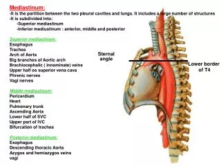
Sternal angle
Mediastinum: It is the partition between the two pleural cavities and lungs. It includes a large number of structures It is subdivided into: -Superior mediastinum -Inferior mediustinum : anterior, middle and posterior Superior mediastinum: Esophagus Trachea Arch of Aorta


Sternal angle
E N D
Presentation Transcript
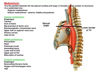
Mediastinum: It is the partition between the two pleural cavities and lungs. It includes a large number of structures It is subdivided into: -Superior mediastinum -Inferior mediustinum : anterior, middle and posterior Superior mediastinum: Esophagus Trachea Arch of Aorta Big branches of Aortic arch Brachiocephalic ( innominate) veins Upper half os superior vena cava Phrenic nerves Vagi nerves Middle mediastinum: Pericardium Heart Pulmonary trunk Ascending Aorta Lower half of SVC Upper part of IVC Bifurcation of trachea Posterior mediastinum: Esophagus Descending thoracic Aorta Azygos and hemiazygos veins vagi ِ Sternal angle angle Lower border of T4
Esophagus: • Length • -begins • Ends • Site and relations • - Blood supply Esophagus Trachea Lt Vagus Rt Vagus • Trachea: • Length • -begins • Ends • Site of trachea and bifurcation) • -Relation • -Main bronchi • . length • . width • . orientation • . divisions
Lt common carotid artery Brachicephalic artery Aorta: 1- Ascending Aorta: Origin, end, branches 2- Arch of aorta: Origin, end, branches 3- descending thoracic Aorta: Origin, end, branches Paired Single Post intercostals pericardial Subcostal mediastinal Phrenic esophageal Broncheal Lt subclavian artery
Brachiocephalic veins: • Comparison between right and left veins: • Begin by union of….. • -length • -end at …. by forming….. • -Tributaries: one of them is internal thoracic vein • Azygos and hemiazygos veins: • Begin at the abdomen. • -end at…… • -Tributaries: • 1- Posterior intercostal • 2- Bronchial • 3- Pericardial • 4- Esophageal • 5- Mediastinal • Superior vena cava : • Begin by union of….. At level of….right 1st costal cartilage • -length • -end at right 3rd costal cartilage by joining the heart • -Tributaries: vena azygos at right 2nd costal cartilage Lt Internal jugular Lt Internal jugular Lt subclavian Lt subclavian Lt subclavian Rt Brachiocephalic. Lt Brachiocephalic Phrenic nerve SVC Vena azygos
Inferior vena cava: -Origin - End Pulmonary Trunk: -Origin -Divisions -Relations
Mediastinum: It is the partition between the two pleural cavities and lungs. It includes a large number of structures It is subdivided into: -Superior mediastinum -Inferior mediustinum : anterior, middle and posterior Superior mediastinum: Esophagus Trachea Arch of Aorta Big branches of Aortic arch Brachiocephalic veins Upper half of superior vena cava Phrenic nerves Vagi nerves Middle mediastinum: Pericardium Heart Pulmonary trunk Ascending Aorta Lower half of SVC Upper part of IVC Bifurcation of trachea Posterior mediastinum: Esophagus Descending thoracic Aorta Azygos and hemiazygos veins vagi ِ Lower border of T4 Sternal angle Esophagus Descending Thoracic Aorta
Lt Subclavian Lt CCA Esophagus Brachiocephalic vein and SVC Trachea Cardiac impression Esophagus Cardiac impression Aorta IVC