Download

1 / 69
700 likes | 1.13k Views
A New Method to Rapidly Evaluate LVEF from a Contractility Polar Map. Lebeau et al. Good afternoon It is my pleasure to present to you a new method to rapidly evaluate LVEF from a contractility polar map in trans thoracic Echocardiography. 2.

E N D
A New Method to Rapidly Evaluate LVEF from a Contractility Polar Map Lebeau et al.
Good afternoon • It is my pleasure to present to you a new method to rapidly evaluate LVEF from a contractility polar map in trans thoracic Echocardiography
2 • This method was developped as a tool to teach cardiology resident and technician how to quantitate LVEF in TTE
Part 1 Part 2 Scientific data supporting LVEF from WMSI A new tool for estimating left ventricular ejection fraction derived from wall motion score index Ref.: Lebeau R. and all, CJC Vol. 19 No 4 March 2003 Semi quantitative visual estimation of LVEF from WMSI map • How to do it • Study and results
3 • The first part of my presentation will concentrate on the scientific data supporting the evaluation of the LVEF from WMSI as published in the CJC in March 2003 • The second part will focus on the visual estimation of the LVEF from the WMSI map, the data behind this rapid method and how to do it
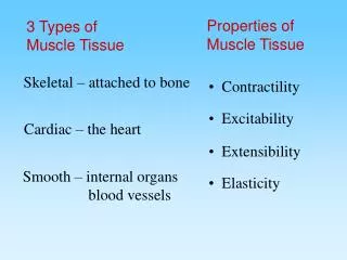
LVEF = systolic function • Commonly use in management of cardiac patients. • Direct correlation between survival and number of hospitalisations post myocardial infarction. • Important in decision making for therapeutics • - Treatment (ICD) • - Cardiac insufficiency • - Post myocardial infarction
4 • The LV systolic function is commonly used in evaluation of cardiac patient • There is a direct correlation between number of hospitalisation post MI, survival and LVEF • Knowledge of LVEF is necessary in therapeutic decision making such as ICD candidate or therapy for heart failure
LVEF / ECHO • 1. Mmode: - Teichholz • - Quinones • 2D : - Planimetric Simpson biplane • - Visual estimate • 3. Doppler and Mmode : Dumesnil • 4. Wall motion score index (Rifkin 1990, r:0.91) • (Berning 1994, r:0.93)
5 • Several methods can be use to assess LVEF, Some using the m-mode such as the Teichholz, Quinones, • Other using 2D data such as the Simpson biplane. and visual estimate. • The doppler and m-mode allow us to use the Dumesnil technique. • As will be shown, the WMSI and the visual method from the WMSI can also be useful
Long axis 4 chambers 2 chambers 4 chambers 2chambers
6 • The polar map is done by evaluating the wall motion in short axis at 3 differents level : the mitral valve, the papillary muscle level and at the apical level. • It can all be done in the short axis view but the long axis view and apical (4C - 2C) can also be used.
7 • This graph represent the 16 segments of the polar map and the coronary blood supply to each segment • The territory supplie by the LAD, by the Cx and by the RCA are described here.
Normal(N) = 1 Hypokinesia(H) = 2 Akinesia(A) = 3 Dyskinesia(D) = 4 Aneuvrysm(AN) = 5 x 8 = 8 x 8 = 24 Total 3216 = 2
8 The WM score is calculated from this polar map by adding all 16 segments. Each segment gets : 1 point if it is normokinetic, 2 point for HK, 3 for AK, 4 for DK and 5 if aneurysmal In this example there are 8 normal segments so we have 8 points, 8 akinetic segments, therefore 24 points . The WMS is therefore 8 + 24 = 32 and the WMSI 32/16 = 2.
Clinical characteristics of the patients Age (years) Sex LV dimension 21 to 88 (mean 69) 140 men 103 women 243 pts Diastolic 40 to 79 (mean 56) Systolic 20 to 70 (mean 42)
9 • In our study, we compared the LVEF mesured by RNA and to the WMSI obtained by echo. • 243 patients were studied, 140 men and 103 women ranging from 21 to 88 years old. • The LV dimension in diastole range from 40 to 79 mm and from 20 to 70 mm in systole. • The polar map by echo and the LVEF in nuclear medicine were obtained within 3 days of each other in all 243 patients.
Methods 150 pts WMSI vs LVEF-RNA Regression Equation: LVEF-RNA = 92,8 – 25,8 * WMSI
Using the LVEF mesured by RNA and the WMSI from the echo, a regression equation was established. This was done with the data from the first 150 patients.
Regression Equation LVEF-RNA = 92,8 – 25,8 x WMSI 92,8-25,8 x 1,0 = 67 (WMS=16) 92,8-25,8 x 1,6 = 53 (WMS=25) 92,8-25,8 x 2,1 = 40 (WMS=33) 92,8-25,8 x 2,6 = 27 (WMS=41) 92,8-25,8 x 2,9 = 15 (WMS=48)
This regression equation allowed us to derive a LVEF from the WMSI and to design this EF table, where each WMSI correspond to a LVEF
Methods 150 pts LVEF -WMSI vs LVEF-RNA Regression Equation: LVEF-RNA = 92,8 – 25,8 * WMSI 243 pts LV function Echo - NM 93pts Regression Equation vs LVEF-RNA 243pts
In the susbsequent 93 patients, the LVEF obtained with regression equation (EF Table) was compared to the RNA LVEF
In the first 150 patients, the correlation between WMSI and RNA EF was 0.82 • The correlation between the LVEF obtained by regression equation, in the subsequent 93 patients and RNA was 0.86 • Correlation for the whole group of 243 patients was 0.88 • If we exclude 20 outliers. (Difference of EF more than 12 % between the two techniques). The correlation between WMSI and RNA EF was 0.92
In various subgroup including patients with dyskinesis (DK), aneurysm (AN), atrial fibrillation (AF), the correlation remains good. Interobserver and intraobserver variability was good.
Teichholz Quinones Simpson Dumesnil LVEF-RNA vs other technics 9pts 10-20 % 9pts 21-30 % 9pts 31-40 % 9pts 41-50 % 9pts 51-60 % 9pts > 61 % 54 pts LVEF-RNA Standard score Score plus : mild HK = 1,5 moderate HK = 2,0 severe HK = 2,5
We compared the WMSI with other techniques in 54 pts with LVEF ranging from 10-61% • We also compared the LVEF by RNA to the LVEF obtained in echo using the Teichholz, Quinones, Simpson,Dumesnil and our score technique. We also use the "score plus" technique where mild HK got 1.5 point, moderate HK 2 points and severe HK 2.5 points
Results Teichholz Quinones Dumesnil Simpson (r = 0,43) (r = 0,50) (r = 0,83) (r = 0,82) WMSI-LVEF vs RNA (54 pts) Standard score (r = 0,85) Score plus (r = 0,83) mild HK = 1,5 moderate HK = 2,0 severe HK = 2,5
The result in 54 patients showed the best correlation with the Simpsons and Dumesnil technique. The correlation with our score (the standard and the modified "score plus") was equally good with a ¨r¨ value of 0.85 and 0.83
Normal(N) = 1 Hypokinesia(H) = 2 Akinesia(A) = 3 Dyskinesia(D) = 4 Aneuvrysm(AN) = 5 x 8 = 8 x 8 = 24 Total 3216 = 2
This is a typical LAD infarct • The WMS score is equal 32 • WMSI is equal 2.0
Estimation of left ventricular ejection fraction according to wall motion score index
The WMSI is 2 and the LVEF is 41% using our regression equation and EF table
16+ Normal(N) = 1 Hypokinesia(H) = 2 Akinesia(A) = 3 Dyskinesia(D) = 4 Aneuvrysm(AN) = 5 x 8 = 8 Normal(N) = 0 Hypokinesia(H) = 1 Akinesia(A) = 2 Dyskinesia(D) = 3 Aneuvrysm(AN) = 4 x 8 = 24 Total 3216 = 2
If we want to use a fast calculation system We begin our calculation at 16 then adding 1 for every HK segment and 2 for every akinetic segments going in a clockwise rotation. The normal segment get no additional points
Fast calculation 16+ 19-20 17-18 23-24 21-22 27-28 25-26 29-30 31-32 Normal(N) = 0 Hypokinesia(H) = 1 Akinesia(A) = 2 Dyskinesia(D) = 3 Aneuvrysm(AN) = 4 Normal(N) = 1 Hypokinesia(H) = 2 Akinesia(A) = 3 Dyskinesia(D) = 4 Aneuvrysm(AN) = 5
The total score is 32 / 16 The LVEF is 41% Try to do a Biplane Simpson in (4C-2C) in 5 seconds.
The goal of our study is to have a visual estimation not dependant of EF table.
Part 2 Semi quantitative visual estimation of LVEF from WMSI map • How to do it • Study and results
I developped a semiquantitative visual estimation of LVEF from the WMSI map even if we don’t do the score calculation
This is done by pattern recognition and it is easy to do and is possible since there is a linear correlation between WMSI and LVEF obtained in nuclear medicine
Estimation of left ventricular ejection fraction according to wall motion score index
This linear correlation is confirm by the EF table. In this table of LVEF we can have 3 subgroup, those with high EF, those with intermediate or low EF The premise behind this rapid method is that if all segments are normal the LVEF is greater or equal to 60% If all are HK, it is 40% If all are AK , it is equal or less than 20%
This drawings represent several possibility For example, if all segment are HK the LVEF is 40% If half of the segments are NK and half HK the LVEF is 50% (mild hypokinesia) If half of the segments are HK and half AK the LVEF is 30% (severe hypokinesia)
Method A + N = H, H (3 + 1) = (2 + 2) D + H = A, A (4 + 2) = (3 + 3) D + N + N = H, H, H AN + N = A, A (5+1) = (3+3) AN + H + H = A, A, A AN + N + N + N = H, H, H, H