Download
1 / 34
360 likes | 1.04k Views
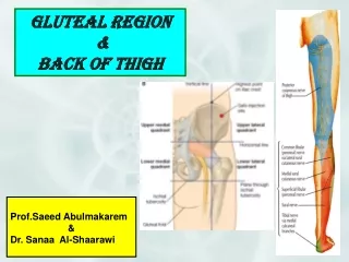
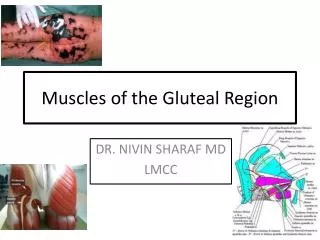
Muscles of the Gluteal Region. DR. NIVIN SHARAF MD LMCC. Objectives.

E N D
Muscles of the Gluteal Region DR. NIVIN SHARAF MD LMCC
Objectives • Identify the bony landmarks of the pelvis and hip on the articulated skeleton and bones. Include: pelvis (ilium, ischium, pubis, iliac crest, iliac fossa, anterior superior iliac spines, pubic tubercle, pubic crest, acetabulum, obturator foramen, greater and lesser sciatic notches, ischial spine, sacroiliac joint, greater and lesser sciatic foramina, ischial tuberosity), femur (head, neck, shaft, greater and lesser trochanters, intertrochanteric line and crest, lineaaspera). • Explain how the anatomical position affects the muscle function. . • Locate the piriformis muscle and the suprapiriform and the infrapiriform spaces. • Identify the superior gluteal nerve in the suprapiriform space and the sciatic and inferior gluteal nerves emerging via infrapiriform space. • . Summarize the muscles of gluteal region and thigh in terms of their location, origin, insertion, nerve supply and actions.
FEMUR Posterior. Anterior. greater trochanter Proximal: -head, -fovea -neck, -greater + lesser trochanters, -intertrochanteric line + crest -gluteal tuberosity -lineaaspera. Distal: -supracondylar lines -epicondyles, -condyles -adductor tubercle head intertrochanteric crest neck lesser trochanter intertrochanteric line gluteal tuberosty linea aspera medial epicondyle lateral epicondyle adductor tubercle supracondylar lines lateral condyle medial condyle
Movements of Hip Joint External rotation Internal rotation
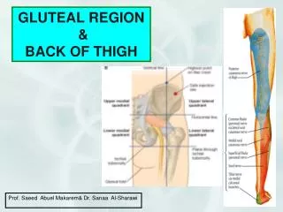
Gluteal region • Gluteal muscles: a-Gluteus maximus b-Gluteus medius c-Gluteus minimus • Tensor fasciae latae -iliotibial tract. • Small lateral rotators of thigh. • Sciatic and posterior cutaneous nerve of thigh. • Superior and inferior gluteal ARTERIES.
Gluteus maximus Tensor Fasciae Latae iliotibial tract Gluteus Maximus gluteus maximus Gluteal region: -Gluteus maximus (most powerful extensor, also lateral rotator) Insertion: Gluteal tuberosity + Iliotibial tract (band) Lateral View Posterior View
Gluteus Maximus FYI Gluteus Maximus and Tensor Fascia Lata insert into Iliotibial Tract - Iliotibial tract is a thickening of the deep fascia (fascia lata) that extends from the ilium to the tibia. - Tension from contraction of gluteus maximus and tensor fasciae latae stabilizes the lower limb as a weight-bearing column.
Tensor Fascia Lata Lateral Posterior
IllioTibial Tract (Band) • Is the thickened lateral part of fascia latae. • Receive insertions of: • 1- tensor fasciae l. • 2- superf ¾ of gluteus maximus. • Attached to oblique ridge on the front of lat condyle of tibia. • Stabilize femur on tibia during standing.
Gluteus Medius Extends, Abducts and Medial and Lateral rotations (Ant and posterior fibers) helps to keep the pelvis level when the opposite leg is raised during activities such as running, Walking, or standing on one leg
Gluteus Minimus Posterior View • Small Lateral Rotators of Thigh • Piriformis. • Obturatorinternus. • Superior gemillus • inferior gemillus • Quadratusfemoris.
Action • Gluteus maximus:1- main extensor of hip. 2- lateral rotation of hip 3- Maintain knee joint in Extension through the iliotibial tract. • gluteus medius, gluteus minimus, tensor fascia latae : 1- extension of hip 2- abduction of hip 3-medial rotation (anterior fibers) 4-contract during walking to prevent tilting of pelvis.
Nerve supply • Inferior gluteal nerve → gluteus maximus. • Superior gluteal nerve →gluteus medius. gluteus minimus. tensor fascia latae
Intrinsic muscles • Infra and supra Piriformis space
Lateral and Medial Rotation of the hip gluteus medius gluteus maximus gluteus minimus Deep to gluteus maximus: -abductors: gluteus medius gluteus minimus (anterior fibres medially rotate) -lateral (external) rotators: piriformis obturator internus (associated gemelli) quadratus femoris [obturator externus is also a lateral rotator] piriformis superior gamellus obturator internus quadratus femoris inferior gamellus
Gluteus medius and minimus: abduction of femur and stabilization of pelvis
Normal Positive sign Trendelenburg Sign. Loss of abductor function (gluteus medius & minimus) causes the pelvis to tilt down when supporting the body on the affected side (*). (I.e. damage to superior gluteal nerve). This function of these muscles is called “stabilization of the pelvis”. *
Superior and Inferior Gluteal Nerves Superior: GluteuMedius Gluteus Minimus Inferior Gluteus Maximus
Intragluteal Injections • What? • Why?
Sciatic Nerve (L4-S3) • Thickestnerve in the body • About 2cm in diameter • L4, L5, S1, S2, and S3 Inside the pelvis • Leave through greater sciatic Foramen, below piriformis.
References • www.netteranatomy.com • www.studentconsult.com • www.google.com • Gray’s Anatomy for students