RISK FACTORS
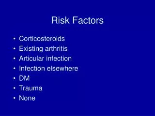
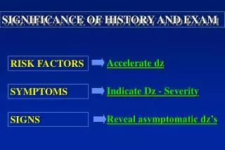
Accelerate dz. Indicate Dz - Severity. Reveal asymptomatic dz’s. SIGNIFICANCE OF HISTORY AND EXAM. RISK FACTORS. SYMPTOMS. SIGNS. Symptoms. PAIN. FUNCTION. ASYMTOMATIC PROBLEMS. HISTORY. EXAM. Risk Factors . Cardiac arrhythmia Hypertension Oral cancer. Signs.

RISK FACTORS
E N D
Presentation Transcript
Accelerate dz Indicate Dz - Severity Reveal asymptomatic dz’s SIGNIFICANCE OF HISTORY AND EXAM RISK FACTORS SYMPTOMS SIGNS
Symptoms PAIN FUNCTION ASYMTOMATIC PROBLEMS HISTORY EXAM Risk Factors Cardiac arrhythmia Hypertension Oral cancer Signs
Ass/Dx (Assessment / Diagnosis) Plan Reassessment EXAM VS (Vital Signs) E/O (Extraoral) I/O (Intraoral) Tests (Vitality/Mobility) RADIOLOGY HISTORY CC (Chief Complaint) HPI (History of present Illness) PDHx (Past Dental History) PMHx (Past Medical History) ROS (Review of System) SHx (Social History) FHx (Family History) Meds (Medications) All (Allergies) STANDARD PATIENT ASSESSMENT
COMPETENCY EXAM • PHYSICAL EXAMINATION • You are required to demonstrate the following diagnostic procedures: • - Take vital signs and document findings in the proper write-up • Palpate regional lymph nodes and landmarks of the neck (hyoid bone, stylohyoid process, thyroid and cricoid cartilage, thyroid gland, mastoid process, vertebral processes, carotids). • Perform a neurologic exam of cranial nerves • Perform regional muscle exam (know origins and insertions of muscles of mastication and major neck muscles) • Perform an intraoral exam, identifying intraoral anatomic landmarks (mucocutaneous junction; labial or lingual frenum; Stenson’s duct; Wharton’s duct; Incisive papilla; Retromolar pad; Maxillary tuberosity; Foliate papillae; Circumvallate papillae; Linea alba; Fordyce granules, if present) • Perform examination of salivary gland function • Know the cardinal features of inflammation • Know how to describe a lesion or a node/lump/mass.
Ass/Dx (Assessment / Diagnosis) Plan Reassessment EXAM VS (Vital Signs) E/O (Extraoral) I/O (Intraoral) Tests (Vitality/Mobility) RADIOLOGY HISTORY CC (Chief Complaint) HPI (History of present Illness) PDHx (Past Dental History) PMHx (Past Medical History) ROS (Review of System) SHx (Social History) FHx (Family History) Meds (Medications) All (Allergies) STANDARD PATIENT ASSESSMENT
VITAL SIGNS: Pulse: 68 reg. BP: 130/85 RAS (right arm sitting)
Ass/Dx (Assessment / Diagnosis) Plan Reassessment EXAM VS (Vital Signs) E/O (Extraoral) I/O (Intraoral) Tests (Vitality/Mobility) RADIOLOGY HISTORY CC (Chief Complaint) HPI (History of present Illness) PDHx (Past Dental History) PMHx (Past Medical History) ROS (Review of System) SHx (Social History) FHx (Family History) Meds (Medications) All (Allergies) STANDARD PATIENT ASSESSMENT
COMPETENCY EXAM • PHYSICAL EXAMINATION • You are required to demonstrate the following diagnostic procedures: • - Take vital signs and document findings in the proper write-up • Palpate regional lymph nodes and landmarks of the neck (hyoid bone, stylohyoid process, thyroid and cricoid cartilage, thyroid gland, mastoid process, vertebral processes, carotids). • Perform a neurologic exam of cranial nerves • Perform regional muscle exam (know origins and insertions of muscles of mastication and major neck muscles) • Perform an intraoral exam, identifying intraoral anatomic landmarks (mucocutaneous junction; labial or lingual frenum; Stenson’s duct; Wharton’s duct; Incisive papilla; Retromolar pad; Maxillary tuberosity; Foliate papillae; Circumvallate papillae; Linea alba; Fordyce granules, if present) • Perform examination of salivary gland function • Know the cardinal features of inflammation • Know how to describe a lesion or a node/lump/mass.
DISASTERS CANCER
4 RF Dz 3 RF 2 RF 1 RF RISK FACTORS (RF)
The concept of T local regional N systemic M TUMOR PAIN local INFECTION
INFECTION NEOPLASM OTHERS • leukemia • connective tissue diseases (e.g. lupus erythematosus) • sarcoidosis CERVICAL LYMPH NODE ENLARGEMENT • bacterial • viral (local / systemic) • fungal • postinfect. fibrosed node • < 40 yrs: lymphoma • > 40 yrs: metastatic SCC
Diagnostic approach for Head and Neck Tumors 1) Rule out lymphadenopathy caused by infection bacterial (odontogenic, tonsils) viral (respiratory epithelium) 2) Search for oral squamous cell carcinoma 3) Follow up in 2 weeks: if still present, refer to ENT specialist for evaluation Asymmetric enlargement of one or more cervical lymph nodes in an adult over 40 is almost always cancerous and usually is due to metastasis from a primary lesion in the mouth or pharynx.
40% stage I & II 60% stage III & IV 80-90% cure 2/3 recurrence < 2 yrs < 1/3 survive 3 yrs. Second primary tumor in successfully treated patients within 5-7 years: up to 40% !!! Follow-up important!!! Importance of early diagnosis of squamous cell carcinoma
therefore……. ...know the H&N anatomy!!! ...performing a H&N exam for EVERY dental check-up!!! ...following up on suspicious signs!!!
Differential diagnosis of lateral tumors of the neck • Salivary gland (sublingual / submandibular / parotis) • Lymph node • Soft tissue tumor (Lipoma, Fibroma) • Neurofibroma • Esophageal diverticulum • Laryngocele • Branchial cyst • Carotid aneurysm • Hyoid bone • Transverse process of cervical spine
Differential diagnosis of midline tumors of the neck • Thyroid • Dermoid cyst • Sebaceous cyst • Thyroglossal duct • Lymphnode
COMPETENCY EXAM • PHYSICAL EXAMINATION • You are required to demonstrate the following diagnostic procedures: • - Take vital signs and document findings in the proper write-up • Palpate regional lymph nodes and landmarks of the neck (hyoid bone, stylohyoid process, thyroid and cricoid cartilage, thyroid gland, mastoid process, vertebral processes, carotids). • Perform a neurologic exam of cranial nerves • Perform regional muscle exam (know origins and insertions of muscles of mastication and major neck muscles) • Perform an intraoral exam, identifying intraoral anatomic landmarks (mucocutaneous junction; labial or lingual frenum; Stenson’s duct; Wharton’s duct; Incisive papilla; Retromolar pad; Maxillary tuberosity; Foliate papillae; Circumvallate papillae; Linea alba; Fordyce granules, if present) • Perform examination of salivary gland function • Know the cardinal features of inflammation • Know how to describe a lesion or a node/lump/mass.
CARDINAL FEATURES OF INFLAMMATION Redness Heat Swelling Pain Dysfunction
COMPETENCY EXAM • PHYSICAL EXAMINATION • You are required to demonstrate the following diagnostic procedures: • - Take vital signs and document findings in the proper write-up • Palpate regional lymph nodes and landmarks of the neck (hyoid bone, stylohyoid process, thyroid and cricoid cartilage, thyroid gland, mastoid process, vertebral processes, carotids). • Perform a neurologic exam of cranial nerves • Perform regional muscle exam (know origins and insertions of muscles of mastication and major neck muscles) • Perform an intraoral exam, identifying intraoral anatomic landmarks (mucocutaneous junction; labial or lingual frenum; Stenson’s duct; Wharton’s duct; Incisive papilla; Retromolar pad; Maxillary tuberosity; Foliate papillae; Circumvallate papillae; Linea alba; Fordyce granules, if present) • Perform examination of salivary gland function • Know the cardinal features of inflammation • Know how to describe a lesion or a node/lump/mass.
DESCRIPTION OF MASSES / LYMPHNODES • Location • Size (< 1cm >) • Tenderness • Consistency • Mobility
COMPETENCY EXAM • PHYSICAL EXAMINATION • You are required to demonstrate the following diagnostic procedures: • - Take vital signs and document findings in the proper write-up • Palpate regional lymph nodes and landmarks of the neck (hyoid bone, stylohyoid process, thyroid and cricoid cartilage, thyroid gland, mastoid process, vertebral processes, carotids). • Perform a neurologic exam of cranial nerves • Perform regional muscle exam (know origins and insertions of muscles of mastication and major neck muscles) • Perform an intraoral exam, identifying intraoral anatomic landmarks (mucocutaneous junction; labial or lingual frenum; Stenson’s duct; Wharton’s duct; Incisive papilla; Retromolar pad; Maxillary tuberosity; Foliate papillae; Circumvallate papillae; Linea alba; Fordyce granules, if present) • Perform examination of salivary gland function • Know the cardinal features of inflammation • Know how to describe a lesion or a node/lump/mass.
COMPETENCY EXAM • PHYSICAL EXAMINATION • You are required to demonstrate the following diagnostic procedures: • - Take vital signs and document findings in the proper write-up • Palpate regional lymph nodes and landmarks of the neck (hyoid bone, stylohyoid process, thyroid and cricoid cartilage, thyroid gland, mastoid process, vertebral processes, carotids). • Perform a neurologic exam of cranial nerves • Perform regional muscle exam (know origins and insertions of muscles of mastication and major neck muscles) • Perform an intraoral exam, identifying intraoral anatomic landmarks (mucocutaneous junction; labial or lingual frenum; Stenson’s duct; Wharton’s duct; Incisive papilla; Retromolar pad; Maxillary tuberosity; Foliate papillae; Circumvallate papillae; Linea alba; Fordyce granules, if present) • Perform examination of salivary gland function • Know the cardinal features of inflammation • Know how to describe a lesion or a node/lump/mass.