Advancements in Dermoscopic Techniques for Skin Cancer Diagnosis
500 likes | 613 Views
The use of dermoscopy, a non-invasive imaging technique, dramatically improves the diagnostic accuracy for skin cancer by up to 30%. Skin cancer, being the most common type of cancer with over one million cases annually, necessitates precise detection methods. This research encompasses the development of a dermoscopic interest point detector and descriptor, leveraging advanced computational techniques to identify critical features from skin lesions. By analyzing dermoscopic images, this work aims to enhance the detection of various skin cancers, including melanoma and carcinoma.

Advancements in Dermoscopic Techniques for Skin Cancer Diagnosis
E N D
Presentation Transcript
1School of Interactive Computing, Georgia Tech • 2Intel Research Pittsburgh Dermoscopic Interest Point Detector and Descriptor Howard Zhou1, Mei Chen2, James M. Rehg1
Skin cancer • Skin cancer : most common type of cancer ( > 1 million ) [ Top 5 categories of estimated annual cancer incidence for 2009 from National Cancer Institute ]
Skin cancer • Skin cancer : most common type of cancer ( > 1 million ) • forms in tissues of the skin Skin lesions [ Image courtesy of “An Atlas of Surface Microscopy of Pigmented Skin Lesions: Dermoscopy” ]
Skin cancer • Skin cancer : most common type of cancer ( > 1 million ) • forms in tissues of the skin Benign lesions Skin cancer [ Image courtesy of “An Atlas of Surface Microscopy of Pigmented Skin Lesions: Dermoscopy” ]
Skin cancer • Skin cancer : most common type of cancer ( > 1 million ) • forms in tissues of the skin Benign lesions Skin cancer Basal cell carcinoma Squamous cell carcinoma Melanoma [ Image courtesy of “An Atlas of Surface Microscopy of Pigmented Skin Lesions: Dermoscopy” ]
Dermoscopy • Non-invasive imaging technique • Improve diagnostic accuracy by 30% Skin cancer Basal cell carcinoma Squamous cell carcinoma Melanoma [ Image courtesy of “An Atlas of Surface Microscopy of Pigmented Skin Lesions: Dermoscopy” ]
Clinical view Dermoscopy • Non-invasive imaging technique • Improve diagnostic accuracy by 30% [ Image courtesy of “An Atlas of Surface Microscopy of Pigmented Skin Lesions: Dermoscopy” ]
Dermoscopy • Non-invasive imaging technique • Improve diagnostic accuracy by 30% • Microscope + light + liquid medium Dermatoscope [ Image courtesy of “An Atlas of Surface Microscopy of Pigmented Skin Lesions: Dermoscopy” ]
Dermoscopy view Dermoscopy • Non-invasive imaging technique • Improve diagnostic accuracy by 30% • Microscope + light + liquid medium • Reveal pigmented structures Dermatoscope [ Image courtesy of “An Atlas of Surface Microscopy of Pigmented Skin Lesions: Dermoscopy” ]
Dermoscopy view Dermoscopic features • Pigmented structures revealed by dermoscopy [ Image courtesy of “An Atlas of Surface Microscopy of Pigmented Skin Lesions: Dermoscopy” ]
Dermoscopy view Dermoscopic features • Pigmented structures revealed by dermoscopy Blue-white veil [ Image courtesy of “An Atlas of Surface Microscopy of Pigmented Skin Lesions: Dermoscopy” ]
Dermoscopy view Dermoscopic features • Pigmented structures revealed by dermoscopy Blue-white veil Scar-like depigmentation [ Image courtesy of “An Atlas of Surface Microscopy of Pigmented Skin Lesions: Dermoscopy” ]
Dermoscopy view Dermoscopic features • Pigmented structures revealed by dermoscopy Blue-white veil Scar-like depigmentation Brown globules [ Image courtesy of “An Atlas of Surface Microscopy of Pigmented Skin Lesions: Dermoscopy” ]
Dermoscopy view Dermoscopic features • Pigmented structures revealed by dermoscopy Blue-white veil Scar-like depigmentation Brown globules Negative network [ Image courtesy of “An Atlas of Surface Microscopy of Pigmented Skin Lesions: Dermoscopy” ]
Dermoscopy view Dermoscopic features • Pigmented structures revealed by dermoscopy • [Betta et al. 2006], [Grana et al. 2006], [Iyatomi et al. 2007],… Blue-white veil Scar-like depigmentation Brown globules Negative network [ Image courtesy of “An Atlas of Surface Microscopy of Pigmented Skin Lesions: Dermoscopy” ]
Dermoscopy view Dermoscopic features • Over 100 dermoscopic features Blue-white veil Scar-like depigmentation Brown globules Negative network … [ Image courtesy of “An Atlas of Surface Microscopy of Pigmented Skin Lesions: Dermoscopy” ]
Dermoscopy view Dermoscopic features • Over 100 dermoscopic features • Multiple binary classifiers for each image BW classifier Blue-white veil Scar-like depigmentation SLD classifier BG classifier Brown globules NN classifier Negative network … … [ Image courtesy of “An Atlas of Surface Microscopy of Pigmented Skin Lesions: Dermoscopy” ]
Dermoscopy view Dermoscopic features • General detector? Blue-white veil Scar-like depigmentation Generalized detector Brown globules Negative network … [ Image courtesy of “An Atlas of Surface Microscopy of Pigmented Skin Lesions: Dermoscopy” ]
Dermoscopy view Dermoscopic features • General detector? Blue-white veil Scar-like depigmentation Generalized detector Brown globules Negative network … • Dermoscopic features consist of low level image characteristics (ridges, blobs, streaks, pigmentation,…) [ Image courtesy of “An Atlas of Surface Microscopy of Pigmented Skin Lesions: Dermoscopy” ]
Dermoscopy view Dermoscopic features • General detector? Blue-white veil Scar-like depigmentation Generalized detector Brown globules Negative network … • Dermoscopic features consist of low level image characteristics (ridges, blobs, streaks, pigmentation,…) • interest points [ Image courtesy of “An Atlas of Surface Microscopy of Pigmented Skin Lesions: Dermoscopy” ]
Dermoscopy view DermoscopicInterest Point (DIP) • General detector: concentration/configuration of interest points • bag-of-visual-words approach Generalized detector Blue-white veil Scar-like depigmentation Brown globules Negative network … • Dermoscopic features consist of low level image characteristics (ridges, blobs, streaks, pigmentation,…) • interest points [ Image courtesy of “An Atlas of Surface Microscopy of Pigmented Skin Lesions: Dermoscopy” ]
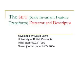
DermoscopicInterest Point (DIP) • Inspired by general interest point detector and descriptors (SIFT & SURF) • We propose Dermoscopic Interest Point (DIP) • detector - to extract these low level building blocks • descriptor – for constructing a general visual vocabulary for dermoscopicfeatures
DermoscopicInterest Point (DIP) • Compared to the general interest point detector and descriptors (SIFT & SURF) • Same key issues • Repeatable • Distinctive • Robust to noiseand deformation (geometric and photometric) • Similar to SIFT & SURF • Corners and blobs • Scale and rotation invariant • In addition • Curvilinear features (fibrillar pattern and radial streaming) • Color component
Detector • Corners and blobs • Fast-Hessian detector [Bay, et al. 2006] Hessian matrix
Detector • Corners and blobs • Fast-Hessian detector [Bay, et al. 2006] • Box filter approximation to replace Gaussian derivatives • Fast using Integral image Hessian matrix
Detector • Corners and blobs • Fast-Hessian detector [Bay, et al. 2006] • Curvilinear structures • Curvilinear detector [Steger, 1996] Hessian matrix
Detector • Corners and blobs • Fast-Hessian detector [Bay, et al. 2006] • Curvilinear structures • Curvilinear detector [Steger, 1996] Hessian matrix
Detector • Corners and blobs • Fast-Hessian detector [Bay, et al. 2006] • Curvilinear structures • Curvilinear detector [Steger, 1996] Hessian matrix
Detector • Corners and blobs • Fast-Hessian detector [Bay, et al. 2006] • Curvilinear structures • Curvilinear detector [Steger, 1996] Hessian matrix
Descriptor • Distinctiveness • Spatially localized information • Distribution of gradient-related features • Dermscopic: color features • Invariance (Repeatability) • Relative strength to reduce the effect of photometric changes • Relative orientation for rotation invariance
Descriptor • Distinctiveness • Spatially localized information • Distribution of gradient-related features • Dermscopic: color features • Invariance (Repeatability) • Relative strength to reduce the effect of photometric changes • Relative orientation for rotation invariance • To construct • Reproducible orientation
Descriptor • Distinctiveness • Spatially localized information • Distribution of gradient-related features • Dermscopic: color features • Invariance (Repeatability) • Relative strength to reduce the effect of photometric changes • Relative orientation for rotation invariance • To construct • Reproducible orientation
Descriptor • Distinctiveness • Spatially localized information • Distribution of gradient-related features • Dermscopic: color features • Invariance (Repeatability) • Relative strength to reduce the effect of photometric changes • Relative orientation for rotation invariance • To construct • Reproducible orientation • Feature vector
Descriptor • Orientation • For rotation invariance • Haar-wavelet responses in x and y direction (in a circular neighborhood)
Descriptor • Orientation • For rotation invariance • Haar-wavelet responses in x and y direction (in a circular neighborhood) • Reponses represented as 2D vectors dy dx
Descriptor • Orientation • For rotation invariance • Haar-wavelet responses in x and y direction (in a circular neighborhood) • Reponses represented as 2D vectors • Average responses in a sliding window of 60 degree dy dx
Descriptor • Orientation • For rotation invariance • Haar-wavelet responses in x and y direction (in a circular neighborhood) • Reponses represented as 2D vectors • Average responses in a sliding window of 60 degree • The longest vector indicates the orientation dy dx
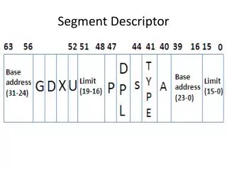
Descriptor • Descriptor components • Context of the descriptor: a square region oriented along the orientation (centered around the interest point) • Local statistics • Uniform 4 x 4 subregions • Intensity gradients (I): Sum of Haar-wavelet responses: dx, dy, |dx|, |dy| • Color statistics (C): Coarse color histogram of the region (alpha & beta channels in L*a*b space) [ Image courtesy of Bay et al. 2006]
Dermoscopy specific • Common interest point descriptor ignores linear features SURF DIP
Conclusion • A generalized framework for characterizing dermoscopic features using Dermoscopic Interest Point (DIP) • A feature detector and a descriptor specifically designed for this purpose • Initial experiments showed that our scheme achieves a comparable level of invariance to lighting, scale, and rotation changes
Future work • Build a vocabulary of dermoscopic features using DIP • Explore the possibility of using DIP in skin CAD related applications: • Dermoscopicfeature extraction and classification • Dermoscopy image registration • Dermoscopy image search and retrieval via dermoscopic features
Acknowledgement • Collaborators (in alphabetical order) • Dr. Laura K. Ferris M.D. Ph.D. UPMC • Richard Gass, Intel Research Pittsburgh • Casey Helfrich, Intel Research Pittsburgh • Many thanks to our anonymous reviewers for their helpful comments and suggestion
Thank you Thank you !
Related publications • Interest pointer detector and descriptors • Distinctive image features from scale-invariant keypointsDavid G. LoweIntl. J. of Computer Vision (IJCV), 2004 • Surf: Speeded up robust featuresHerbert Bay, Tinnetuytelaars, and Luc Van Gool,in Eur. Conf. on Computer Vision (ECCV),2006 • An unbiased detector of curvilinear structuresCarsten Steger,IEEE Trans. Pattern Anal. Machine Intell.(PAMI) 1996
Outline • Introduction • Detector • Corners and blobs • Curvilinear structures • Descriptor • Orientation • Descriptor components • Validation • Conclusion
Dermoscopic features • A Pigmented Skin Lesion (PSL) typically has several dermoscopic features • Over 100 of these features
n(x) Detecting line points Cross section Curve L’ = 0 L’’ large n(x) L(x) [ Steger 1998, ”An Unbiased Detector of Curvilinear Structures” ]