Download
1 / 24
240 likes | 350 Views
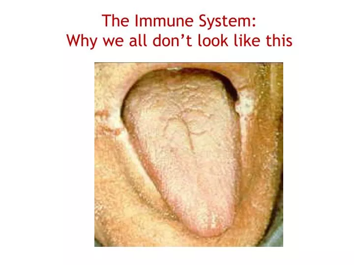
The Immune System: Why we all don’t look like this. I. Sources of Immunity A. Inherited or Acquired Inherited (aka innate or inborn)- from development in the womb Acquired- after birth, from body's exposure to an antigen (NATURAL) or immunization (ARTIFICIAL). (Types of Acquired Immunities)

E N D
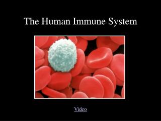
The Immune System: Why we all don’t look like this
I. Sources of Immunity • A. Inherited or Acquired • Inherited (aka innate or inborn)- from development in the womb • Acquired- after birth, from body'sexposure to an antigen (NATURAL) orimmunization (ARTIFICIAL)
(Types of Acquired Immunities) a. Active- own body's response to antigenby making antibody, longer-lasting ex. Measles (nat.), Polio vacc. (artif.) b. Passive- body is given the antibodies directly, temporary but immediate ex. Breast feeding (nat) vs
II. Nonspecific defense mechanisms: general, innatebarriers to infection A. Mechanical and Chemical Barriers- 1st line 1. Skin and mucosa 2. Secretions like sebum, mucus, enzymes Genetically Modified Skin that is resistant to infection
B. Inflammation- 2nd line • Mediators released after tissue damage histamine, kinins, prostaglandins • Redness, heat, swelling, pain, vasodilation,fever (mild fever good) • Chemotaxis- mediators attract more WBC
C. Phagocytosis- part of 2nd line- ingestion of microorganisms; pseudopods surround and lysosomes digest
Trypanosoma : human parasite that causes African sleeping sickness, transmitted by tsetse flies
C. • Neutrophil- granular WBC, mostnumerous, form pus when die • Macrophage- aka monocyte, agranularwbc, names differ based on location
D. Natural Killer Cells- part of 2nd line 1. Police blood and lymph 2. Lymphocytes lyse tumor cells or viruses
E. Interferon- part of 2nd line 1. Protein that interferes with reproductionof viruses and cancer 2. 3 kinds: leukocyte a, fibroblast b,immune g 3. Genetic engineering IFN -2a is indicated for hairy cell leukemia (HCL), acquired immune deficiency syndrome (AIDS)-related Kaposi's sarcoma (KS), chronic-phase Philadelphia (Ph) chromosome-positive chronic myelogenous leukemia (CML) and chronic hepatitis C (CHC).
F. Complements- 2nd line • 20 inactive plasms proteins or enzymesthat are triggered by pathogens • cause lysis by binding to pathogen'ssurface • Increased complement in: • Cancer • Certain infections • Ulcerative colitis • Decreased complement in: • Cirrhosis • Glomerulonephritis • Hereditary angioedema • Hepatitis • Kidney transplant rejection • Malnutrition
Tissue macrophages (pink/purple), T lymphocytes (green), and human red blood cells from a leg wound. • A tissue macrophage (pink) is a mature phagocyte that can ingest and destroy invading microbes, foreign particles and cellular debris. • A monocyte (purple)is a circulating phagocyte that ingests microbes, invading particles, and cellular debris. • Lymphocytes are involved in the specific immune response • Precursor T cells (T lymphocytes) • Migrating to the thymus where they develop into specialized cells (helper T and killer T cells) that are able to identify antigens and infected tissue cells • Precursor B cells (B lymphocytes)
III. Specific Mechanisms- fight specific invaders;3rd line Lymphocytes- develop in red blood marrow,fetal liver, lymph nodes, thymus, and spleen
B. Antigens aka immunogens- foreign invaders or cancer cells epitope identifies antigen Epitope is what the antibody recognizes
C. Antibodies aka immunoglobulins (Ig)- • protein made by B cells • containsvariable or binding site to join withepitope of antigen
1. Structure a. Y-shaped b. contains 4 polypeptide chains: 2 heavy & 2 light c. variable region differs d. 5 classes: IgM, IgA, IgD, IgE, IgG
2. Functions of antibodies a. forms antigen-antibody complex b. inactivates antigens c. allows macrophages to destroy thecomplex d. changes shape so complement canlysis e. initiates release of mediators
A. Cell-mediated Immunity (T cells) • Have surface receptors that match antigen's epitope • When activate, secrete lymphokines or cytokines to promote phagocytosis or release lymphotoxins. • Killer T cells- release lymphotoxin; kill cells taken over by virus or cancer • Helper T cells- help B cells activate into plasma cells to make antibodies • Suppressor T cells- stop T (and B) cells from activating
B. Antibody-mediated immunity, aka humoral 1. Inactivated B cells become activated when in contact with epitope of antigen 2. B cells can generate 2000 antibodies/ sec 3. Memory B cells remain and becomeactivated later
V. Organ Transplants and Rejections • A. Common since 1970s • B. Must check ABO groups • C. Post-operation treatment w/immunosuppressive therapy • D. 4 kinds • autograph- same person • isograph- identical twins • allograph- same species • xenograph- different species