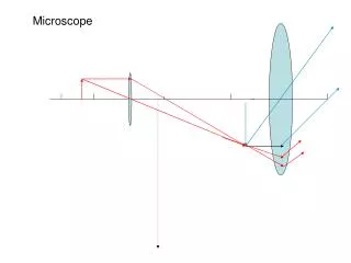
Microscope Imaging Processing
Microscope Imaging Processing. By Jacob, Eric, Woo Y eon , Alana. Definition. Microscope Image Processing is a technique in which data is digitized and various mathematical operations are applied to the data.

Microscope Imaging Processing
E N D
Presentation Transcript
Microscope Imaging Processing By Jacob, Eric, Woo Yeon, Alana
Definition • Microscope Image Processing is a technique in which data is digitized and various mathematical operations are applied to the data. • It is generally done with a digital computer, in order to create an enhanced image that is more useful or pleasing to a human observer. • Microscope Image Processing is also known as picture processing.
When and where is it useful? • Microscope Imaging Process is useful in electrical engineering and computer science. • It is helpful to enhance/reconstruct images. • It allows a much wider range of algorithms (a step-by-step problem-solving procedure, solving a problem in a finite number of steps) to be applied to the input data and can avoid problems such as the build-up of noise and signal distortion during processing.
Hardware • Tools: Digital Camera, Microscope, Computer, and an imaging editing software.
Process • 1st- Strap the digital camera on the microscope • Camera needs to produce less noise because microscopes have an intrinsic limiting resolution • Larger pixels preferred • 2nd – Take the picture • 3rd- Edit the picture • Image is most likely has low contrast • The more bits the better • 4th- store the picture • Some bits will be lost
Object of Interest • Microscopic objects • Animals • Used in medicine, biological research, drug testing, etc.
Software • Netpbm • Image Magick • GIMP This is an enormous set of command line programs that can be used to interconvert virtually any common image formats, and can also perform certain image processing functions. This package has no provision for viewing images. This package can read many image formats, and perform several types of image processing functions. It has a very useful image display program called "display." It can read and write formats that have 16 bits/color. There are at least two separate versions of this package being maintained at this point. This program has been around for several years and is very stable. It has many extraordinary capabilities. Unfortunately it cannot read formats with more than 8 bits/color. It takes "plug ins" that can add more capabilities than it already has. It is somewhat user hostile, and the documentation is not as great as it could be.
Example of techniques • Point to point operation : subtraction, mapping • Statistical operation : histogram • Spatial filtering operation : convolution • Morphological operation : erosion, dilation • Geometric transformation operation : resize
Steps of the operation Decomposing the image green red blue
Low contrast the pixel values range from 0 to the image's specified maximum High contrast
Example • Microscope imaging processing can greatly increase the detail of pictures taken with a microscope. For example, when looking at bacteria, an image is vague unless it’s touched up with image processing. In this image, contrast was added to yeast cells, and colors were added to separate different parts of the cells. This example distinguishes the cell’s nucleus in blue, and other areas in green, with black background to highlight the sides of the cell. • The importance of this kind of processing is to give people in this field a better understanding of what the cells they’re working with look like. Scientists can target a cell’s cell wall to remove parts of it and also the nucleus to inject chemicals. They could also observe what parts of the cell changes when a yeast cell divides. Scientists can label cells with fluorescent light which allows them to delicately observe molecules in a cell’s body.
Applications • Other applications of image processing in Microscope Images include establishing distance inside the picture, and averaging pictures to minimize the blur in the picture.