Download

1 / 31
320 likes | 334 Views
Learn the key elements of liver palpation, spleen examination, gallbladder assessment, kidney palpation, and more. Understand normal findings, abnormalities, and diagnostic signs. Master the palpation of abdominal masses, fluid wave thrill, and succussion splash.

E N D
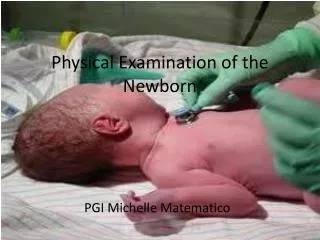
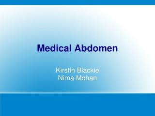
The Physical Examination of Abdomen2nd Affiliated Hospital China Medical University
The contents of palpation1.abdominal muscles tensity 2.tenderness and rebound tenderness 3.abdominal organs4.abdominal masses 5.fluid thrill 6.succussion splash
When you palpate the liver you should pay special attention to the following items (1) size (2) consistency (3) contour margin (4) tenderness (5) pulsation (6) friction sound (7) hepatojugulor reflux (8) liver thrill
(1). The size of liverin healthy person the liver is not palpable or palpable within 1 cm below the costal margin 3 cm below the xiphoid hepatomegaly diffuse hepatitis fatty liver early cirrhosis of liver hepatic engorgement淤血
located enlargement of liver hepatic cyst hepatic abscess shrinking of liver acute liver necrosis cirrhosis of liver hepatometry midclavicular line how many cm below costal margin abdominal middle line how many cm below xiphoid process
(2)Consistency the consistency of liver is divided into 3 degrees soft as like lips seen in normal liver middle hard as like nose acute chronic hepatitis hard as like frontalis cirrhosis carcinoma fluctuation big surface cyst
(3) Contour and margin normal liver: the surface is smooth and margin is regular irregular nodular dull: cancer (4) tenderness normal liver: no tenderness light: hepatitis sever: hepatic abscess
(5) Pulsationnormal liver: no pulsation distensible pulsation: tricuspid valve incompetencetransmitted pulsation: aneurysm (6) friction sensation area: perihepatitis (7) hepatojugulor reflux(8) liver thrill: echinococcosis
The error of palpation(1) patient can`t coordinate with doctor (2) massive liver to palpate over liver (3) the doctor`s hand presses too heavy to move liver down
(4) some organs may be misapprehended the liver such as: transverse colon lower extreme of right kidney tendon of abdominal rectus
2). Palpation of spleenthe position of the patient supine right lateral decubitus palpating methods palpation with single hand bimanual palpation ballottement
Splenometry1line (A-B line) midclavicular line 2line (A-C line) the longest line 3 line (D-F line)
Splenomegalylight <2 cm chronic hepatitis, typhoid fever, middle 2 cm – umbilicus cirrhosis of liver chronic hemolytic jaundice heavy below umbilicus chronic granulocytic leukemia myelofibrosis
3).Palpation of the gallbladder Murphy’s sign: acute cholecystitis Courvoisier’s sign pacreatic carcinoma
4). Palpation of kidneybimanual palpation to palpate right kidney
Tenderness pointskidney urinary tube point (1) upper ureter point (2) middle ureter point ureteritis ureterolithiasis (3) costovertebral(4) costolumber pyelonephritis TB kidney pyelolithiasis
4. Palpation of massesthe masses of abdomen may be caused by enlarged organ ectopic organ cyst carcinoma inflammatory tissues enlarged lymphnode
(1)Normal masses of abdomentendon of abdominal rectus lumber vertebral body sacral promontory sigmoid colon transverse colon cecum
(2).Abnormal mass of abdomenwhen you palpate the mass of abdomen you should describe the location size contour consistency tenderness mobility pulsation
5. Fluid wave thrillwith the patient in supine position, the examiner’s left hand is placed on the patient’ s right flank, an assistant (another person) places one hand on the middle of the abdomen to prevent the transmission of any wave through the tissues of the abdominal wall
Succussion splashthis examining method can check for retention of gastric fluid. If succussion splash is positive after meal 6-8 hours indicating pyloric obstruction