Download

1 / 31
340 likes | 492 Views
Unit Title: Excitable Cells Tidbit Title: Understanding membrane potentia l. Howard Sirotkin, Abhay Deshpande: Stony Brook University Thomas Abbott, Kristen Kimball: UCONN Jagan Srinivasan, Elizabeth Ryder: WPI Neurobiology & Physiology Group Northeast Summer Institute

E N D
Unit Title: Excitable CellsTidbit Title: Understanding membrane potential Howard Sirotkin, Abhay Deshpande: Stony Brook University Thomas Abbott, Kristen Kimball: UCONN Jagan Srinivasan, Elizabeth Ryder: WPI Neurobiology & Physiology Group Northeast Summer Institute Stony Brook University 2013 Facilitators: Thomas Torello, Harvard University Cynthia Wagner, UMBC
Materials required for this activity: • A large sheet of white paper (approximately 3’ x 4’). Note: this entire activity can be done with smaller activity boards. • Sticky notes (3 colors) and sizes (small and large). • Prepare the activity boards: • Draw a representation of the phospholipid bilayer on the white paper across the long axis (“landscape mode”). The bilayer should divide the paper in half. • Using small stickies, prepare sticky note “ions” (Na+ and K+). These individual ions represent the ions that can move across the membrane to establish the electrical gradient). One color sticky will be K+ ions, the other will be Na+ ions (e.g. pink stickies for K+ ions and green stickies Na+ ions). Label the sticky notes (6-10 per activity board). • Using larger stickies, prepare “pools” of immovable K+ and Na+ ions (these represent the intracellular and extracellular excess concentrations of ions that maintain the chemical concentration). Ensure that the color is consistent between the big and small stickies (e.g. pink stickies for K+ ions and green stickies Na+ ions). • Using the third color stickies, label the small stickies ‘-’, and the large stickies will be ‘pools’ of – charges. These ‘-’ charges are kept deliberately vague, as they might represent Cl-, or other larger anions. • Using larger stickies, prepare Na+ and K+ channels. Ensure that the color is consistent between the big and small stickies (e.g. pink stickies for K+ channels and green stickies Na+ channels).
Context of our tidbit • Introductory Biology Course /Applications of Physics to Biology • Beginning of a Neurophysiology Section of a advanced Animal Physiology course • Students are already familiar with: • Cell membrane structure and function • The chemical and structural nature of neurons
Tidbit: Understanding Membrane Potential Goals for the Tidbit: • To understand membrane potential, how it is generated and why it matters • To recognize the interdisciplinary nature of neurobiology
Objectives for the tidbit Students will be able to diagram how chemical and electrical gradients generate membrane potential
Different activities require different types of neurons… ….but all neurons need to have a baseline resting potential in order to be excitable.
From your pretest… • Which of the following describes the ion concentrations in a typical neuron? A) [Na+] high inside, [K+] high inside B) [Na+] high outside, [K+] high outside C) [Na+] high inside, [K+] high outside D) [Na+] high outside, [K+] high inside
Set up your activity sheet using what you know about the cell
OUT Na+ Na+ Na+ Na+ Na+ Na+ Na+ Na+ Na+ Na+ Na+ K+ K+ K+ K+ K+ K+ K+ IN K+ K+ K+ K+
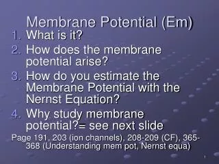
Membrane Potential • The potential difference across the membrane, or membrane potential (Vm), is simply the difference in charge between the inside of the cell and the outside of the cell. • Potential difference is measured in volts; for cells the difference is thousandths of a volt, therefore "millivolt“ (mV) is the unit. • By convention, we state the membrane potential as INSIDE relative to OUTSIDE the membrane. Thus, if Vm = -50 mV, the inside of the cell is more negatively charged than the outside.
What is the membrane potential (Vm) across this membrane? • < 0 mV (more negative inside) • 0 mV (neutral) • > 0 mV (more positive inside)
OUT Na+ Na+ Na+ Na+ Na+ Na+ Na+ Na+ Na+ Na+ Na+ K+ K+ K+ K+ K+ K+ K+ K+ Leak channels K+ K+ K+ K+ IN K+ K+ K+ K+
OUT Let’s start by considering only the K+ ions. K+ K+ K+ K+ K+ K+ K+ K+ Leak channels What happens now ?? K+ K+ K+ K+ IN K+ K+ K+ K+
Which of these pictures is the best representation of what happens? B A C Tiny numbers of K+ cross membrane No anion channels in membrane
CHEMICAL force driving K+ out of the cell is equal toELECTRICAL force driving K+ into the cell. This membrane potential is called the equilibrium potential for K+, or EK. Vm = EK when only K+ ion channels are present. Are K+ ions still moving?
So, what is the Vm across this membrane? • < 0 mV (more negative inside) • 0 mV (Neutral) • > 0 mV (more positive inside)
OUT Na+ Na+ Na+ Na+ Na+ Na+ Na+ Na+ Now let’s look at Na+ ions. Na+ Na+ Na+ Na+ Na+ Na+ Na+ Na+ Leak channels What happens now ?? IN
What is the Vm across this membrane? • < 0 mV (more negative inside) • 0 mV (neutral) • > 0 mV (more positive inside)
OUT Na+ Na+ Na+ Na+ Na+ Na+ Na+ Na+ Na+ Na+ Na+ Na+ Na+ Na+ Na+ Does your sheet look something like this? When only Na+ channels are open in the membrane, Vm = ???? IN
Now let’s look at a slightly more realistic representation of the resting neuron, incorporating both K+ and Na+ ions and channels
OUT Na+ Na+ Na+ Na+ Na+ Na+ Na+ Na+ Na+ Na+ Na+ Na+ K+ K+ K+ K+ K+ K+ K+ What happens now ?? K+ K+ K+ K+ IN K+ K+ K+ K+
Compared with a membrane containing only K+ channels, the Vm of the resting cell is • More negative than EK • Equal to EK • More positive than EK
Basic physics in understanding cell functionMembrane Capacitor circuit CURRENT “I” BATTERY “V” volts Na+ Na+ Na+ Na+ Na+ Na+ Na+ + + + + + + + + + + + + + + + CAPACITOR “C” K+ K+ − − − − − − − − − − − − − − − + + + + + + + + + + + + + + + + + + + + + + + + RESISTANCE “R” Ohms − − − − − − − − − − − − − − − − − − − − − − − - K+ K+ K+ K+ K+ Channel Conductance measured in ‘G’ Seimens G = 1/R
Summary • Concentration gradients across membranes containing selective ion channels establish membrane potentials • The equilibrium potential for an ion Eion occurs when the concentration gradient ‘pushing’ the ion in one direction is balanced by the electrical gradient ‘pulling’ the ion in the opposite direction • The resting potential in a cell results from opposing Na+ and K+ concentration gradients, combined with the presence of both K+ and Na+ leak ion channels • Cell membranes can be represented by electrical circuits
In the next class, we will look at how changes in membrane potential create action potentials and how they are propagated…..
Homework We talked about several situations in class today: a membrane with only K+ channels, a membrane with only Na+ channels, and a resting cell membrane with a mixture of channels. • How does the membrane potential differ in each of these situations? • Are Na+ and K+ ions flowing in the resting cell that has a mixture of channels? If so, in what directions?