Download
1 / 24
250 likes | 507 Views
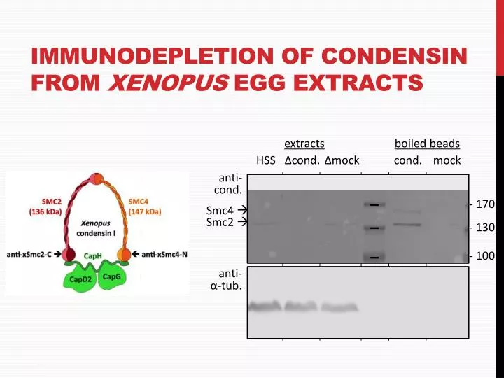
Immunodepletion of Condensin from Xenopus Egg Extracts. extracts. boiled beads. HSS. Δcond . Δmock. cond. mock. anti- cond . - 170. Smc4 . Smc2 . - 130. - 100. anti- α- tub . Fluorescence microscopy: Visualising fitc -stained samples. Camera/Eyepiece.

E N D
Immunodepletion of Condensinfrom Xenopus Egg Extracts extracts boiled beads HSS Δcond. Δmock cond. mock anti- cond. - 170 Smc4 Smc2 - 130 - 100 anti- α-tub.
Fluorescence microscopy: Visualisingfitc-stained samples Camera/Eyepiece Emission filter (525nm+/-25 nm) Excitation filter (488nm+/-20 nm) Dichroic beam splitter(>495 nm)
Fluorescence microscopy:Microscope set-up Emission filter Excitation filter Beam splitter
Example: Bright-field microscopy of a stained sample Kidney ducts stained with hematoxylin (blue, basic extracellular matrix) and eosin (pink, acidic nuclei) Source: MBC
Bright-field microscopy • Based on differential absorption of light by objects • Absorption: Decrease in the amplitude of a light wave (i.e. object gets darker) • Absorption may be wavelength-independent or wavelength-specific (e.g. chloroplasts are green under the microscope, the amplitude of all other wavelengths is reduced) • Objects visible by bright-field microscopy are called “amplitude objects”
Phase-contrast microscopy • Thin objects (e.g. single cells) don’t absorb sufficient light to be good “amplitude objects” • However, all objects shift the phase of a passing light-beam by a fraction of their wavelength. They are called “phase objects”. • Using special optics this (invisible) phase shift can be converted into a (visible) amplitude shift • This conversion is based in interference between the direct light beam and the phase-shifted light beam
Light path in phase contrast microscopy Δ1/4λ Δ1/4λ -1/4λ on the diffracted beam (passing through the retarder of the phase ring) Direct beam Diffracted beam -1/4λ on the diffracted beam (passing through the specimen) Net result: shift of 1/2λ of the diffracted beam results in negative interference between direct and diffracted beam apparent conversion of a “phase object” into an “amplitude object”
Types of light microscopy Bright-field Phase-contrast Differential-interference contrast (DIC) Source: MBC
Principle of fluorescence • Fluorochromes can be excited by a particular wavelength and emit light of a longer wavelength Stokes shift. =heat
Probe detection • Antibodies or nucleic acid probes can be conjugated to fluorescent dyes, such as FITC (fluorescein-isothiocyanate) Fluorescent group Reactive group for conjugation to other molecules via amine groups
Scale Bars 200 µm • all microscopicimages must have a scale bar • experimental determinationofscale bar: takeimageofhemocytometerwithsquaresofknowndimensions (e.g. Thoma) • calculatelengthfrompixelasoutlinedbelow:
Calculating magnification for digital microscopy • Pixel size: 6.8 µm • CCD chip dimension: 1360 x 1024 pixel • Microscope magnification: 100x • 6.8 µm/100 x1360=92.48 µm • 6.8 µm/100 x 1024=69.63 µm • One image is 92.48 µm in length and 69.63 µm in height
Actin fibres in interphase cells 10 µm Stained with Phalloidin-Fluorescein DNA (DAPI stain) pseudocoloured in red
Microtubules in interphase cell 10 µm Stained with anti-tubulin antibodies and secondary fluorescein antibodies DNA (DAPI stain) pseudocoloured in red
Macrophage phagocytosis: Signaling through heterotrimeric g-proteins
Macrophage phagocytosis: Chemokines act through heterotrimeric g-proteins Artificially activated by phorbol ester (mimics DAG) Artificially elevated by ionomycin
Macrophage phagocytosis: E. Coli lipopolysaccharide (LPS) Lipid A
Lymphocyte proliferation • Concanavalin A: • Polyvalent lectin • α-D-mannosyl and α-D-glycosyl binding • Mitogen • Polyclonal activation (in contrast to antigen-mediated clonal expansion) • Pleiotropic effects • Metabolic stimulation • Receptor clustering (lipid raft)?