The cell Nucleus
Chapter 9. The cell Nucleus. Biology Department of the Basic Teaching Colledge. Xiamixinuer · Yilike. The May of 2012. Chapter 9 Nucleus. Learning Objectives

The cell Nucleus
E N D
Presentation Transcript
Chapter 9 The cell Nucleus Biology Department of the Basic Teaching Colledge Xiamixinuer·Yilike The May of 2012
Chapter 9Nucleus Learning Objectives 1. Mastering: ultrastructure of nuclear envelop; nuclear pore complex; composition and four levels organization of chromatin; packaging of chromatin; types of chromatin. 2. Comprehending: structure and function of nucleolus; function of nuclear pore complex; process of RNA processing. 3. Understanding: basic function of nucleus; nuclear matrix.
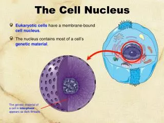
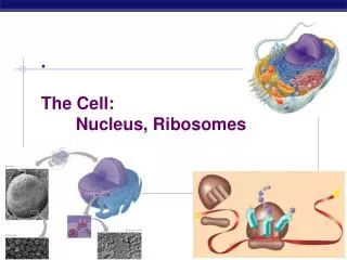
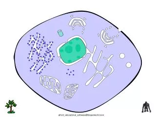
1. The nucleus: Nuclear envelope and NPC A. Structure: Double-membrane nuclear envelope surrounds the nucleus Structure of the interphase nucleus
The main functions of nucleus 1. Carry genetic information(DNA); 2.Duplicate,transcript of genetic information and control protein synthesis; 3.Regulation and control centre of living action of cells.
NP(nucleoplasmic index) • NP= Vn/(Vc-Vn) • Usually size of nucleus can be estimated by calculate the NP. • Normal cell NP≈0.5, • Dividing cell NP>0.5, • Aging cell NP<0.5。
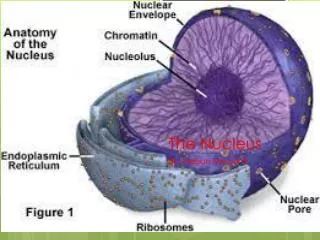
A typical nonmitotic nucleus includes four major components. nuclear envelope interphase nucleus nucleoluos chromatin matrix
B. The nuclear envelope consists of two membranes by a perinuclear space.
The inner surface of the nuclear envelope is lined by the nuclear lamina • The nuclear lamina supports the nuclear envelope: Gives shape and stability of nuclear envelope; Provides a structure link between chromatin and nuclear envelope; • The nuclear lamina is composed of lamins. • The integrity of the nuclear lamina is regulated by phosphorylation and dephosphorylation.
Breakdown and reformation of nuclear envelope during mitosis
The structures of nuclear envelope outer nuclear membrane inner nuclear membrane perinuclear space nuclear pores nuclear lamina
Ring subunit:8 pair Annularsubunit:8 Old model structure Central plug:1 fibril:
outer membrane Perinuclear space pore Inner membrane 蛋 B B B B B B B A A A A A A A C C C C C C C nuclear lamina lamina A.B.C
D. Molecules enter and exit the nucleus through nuclear pore complex Bidirectional traffic
Molecules enter and exit the nucleus through nuclear pore complex
3000-4000 NPC/cell(mammalian); To import about 106 histone/3 mins.(DNA-sythesizing cell) = 100 histone/ min/NPC • Each NPC contains one or more open aqueous channels: 9 nm in diameter and 15 nm long • The effective size of these channels has been determined by injecting various sizes of colloidal gold particles and examined by electron microscopy. • <10 nm in diameter • <60kd globular protein • Passive transport—passively diffuse Able to enter the nucleus
Active transport • Transport of large proteins into nucleus needs nuclear localization signal (NLS) Pore diameter is about 9nm but much larger objects, including ribosomal subunits can pass through (albeit slower than smaller molecules). This implies that recognition of appropriate signal allows temporary stretching of the pore. Energy is required.
Chapter 9 section 2 Nucleolus and Chromosomes • Learning Objectives • The components of chromatin and packaging of chromosome • (2) Nucleolus.
Chromatin fiber human lymphocyte nucleus under SEM human nucleus under LM
Eukaryotes package DNA in Chromatin and chromosomes • Each human cell contains about 2 m of DNA within nucleus if stretched end-to-end, yet the nucleus of a human cell itself is only about 6 μm in diameter. • Compaction ratio=nearly 10000-fold. • (Chromosome 22: DNA 1.5cm2 μ m)
Chromosomes exist in different states throughout the life of a cell. Chromatin: (Interphase) Fibers, 10-30nm in diameter,dispersed through the nucleus. DNA+Proteins+non-Proteins+RNA. Chromosomes: (M phase) Cell division, these fibers condense and fold into larger, compact structure.
Levels of organization of chromatin
B. Nucleosomes are packed together to form chromatin fibers and chromosomes • 4 degree folding model Nonhistone proteins provide a structural scaffold for long chromatin loops. 2nm DNA –11nm nucleosome – 30nm fiber – Loop– Metaphase chromosome
4 degree folding model • First degree: Nucleosomes • Second degree: Filament 10nm in diameter • Third level: Fiber,30nm in diameter • Fourth level: Chromosome
A. DNA packaging:First degree of packaging is Nucleosomes Nucleosomes are the basic unit of chromatin structure.
Evidence: (1)Electron micrographs of chromatin fibers Isolated from interphase nucleus: 30nm thick Chromatin unpacked, show the nuclesome
Evidence: (3)X-ray diffraction studies 3-D of nucleosome Nature 389:251, 1997
Digested briefly: H1+Octamer+200bpDNA Digested longer: Octamer+146bp H1 is released Core particle Structure of nucleosome
NUCLEOSOME H3 H3 H4 H4 H1 H3 H3 H4 H4 10nm H2B H2A H2A H1 H2B CORE OF PERIPHERAL HISTON H2B DNA (146bp、1.75 ROUND) H2A H2A HISTONE:2(H2A、H2B、H3、H4)OCTAMER H2B CORE LINKER DNA(60bp) DNA MOLECULE:146bp、1.75 ROUND NUCLEOSOME HISTONE:H1 LINKER DNA MOLECULE:60bp
Histone octamer: • (H2A-H2B)-(H3-H4)-(H3-H4)-(H2A-H2B) • A histone octamer forms the nucleosome core • Where is the histone H1? • H1 molecules are associated with the linker region. 146+15~50bp linker DNA • 200bpDNA: Linker DNA:15-50bp Nucleosomal DNA:146bp to wrap 1.75 times around the histone core.
Evidence: (2)Nuclease digestion (Rat liver chromatin) The basic repeat unit, containing an average of 200bp of DNA associated with a protein particle, is the nucleosome
Histones: The most abundant proteins associated with eukaryotic DNA Rich in positively charged basic amino acids, which interact with the negatively charged phosphate groups in DNA The amino acid sequences of histones H2A, H2B, H3, H4 are remarkably similar among distantly species
Third level: fiber,30nm in diameter • Fourth level: chromosome
B. Euchromatin and Heterochromatin • Euchromatin • The possibility of transcription; • The types of chromosomal structure—30-nm fibers and looped domains; • Light-staining, less condensed; • Transcriptional activity
Heterochromatin: • Dark-staining, condensed chromatin; • No transcriptional activity; in a typical mammalian cell, approximately 10% of the genome is packaged into heterochromatins forming CEN and TEL • Divided into two classes: • Constitutive & facultative Example of facultative heterochromatin: Random inactivation of X chromosome in different cells during early embryonic development Inactivated at certain phase of life Compacted state at all time: Centromere Barr body in a woman’s cell
3. Chromosome number,size,and shape at metaphase are species specific Karyotype Banding Chromatids
Main structures of chromosome: Including centromere, arm (p and q), secondary constriction, telomere and satellite. • Centromere & Kinetochore Centromere: Highly repeated DNA+Kinetochore C. Main structures of chromosome • The Centromere and Kinetochore: serve as a site for the attachment of spindle microtubules during mitosis and meiosis The structure of a human CEN
A typical mitotic chromosome at metaphase satellite secondary constriction nucleolar organizing region,NOR centromere telomere Human chromosome No.14
Three functional elements are required for replication and stable inheritance of chromosomes