Download
1 / 68
680 likes | 729 Views
Explore the components of hemostasis, vasculature, coagulation proteins, and platelet functions in the context of clot formation, dissolution, and blood vessel integrity. Learn about platelet activation pathways and the role of various factors and inhibitors in regulating coagulation. Gain insights into the intrinsic and extrinsic pathways of the coagulation cascade and the impact of abnormal hemostasis on thrombus formation and bleeding tendencies.

E N D
Introduction to Special Coagulation Ahmad Sh. Silmi Hematology Lecturer Medical Tech. Dept IUG
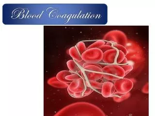
Hemostasis • Hemostasis: The balance between clotting and bleeding • Components of Hemostasis: • Vasculature • Coagulation proteins • Platelets Clot Formation Clot Dissolution
Blood Vessels • Intact endothelium forms a thromboresistant surface • endothelial cells have additional synthetic functions.
Prostacyclin Thromboxane A2 ELAMs, ICAMs von Willebrand factor Vascular Endothelium Function Vasodilation, inhibition of platelet aggregation From platelets, constrict muscular arteries Cytokines induce synthesis to promote leukocyte adhesion Promote platelet-collagen adhesion to exposed sub-endothelium
Tissue factor pathway inhibitor Thrombomodulin Tissue plasminogen activator Heparan sulfate proteoglycans Tissue factor Anticoagulant- Inhibits coagulation extrinsic pathway Anticoagulant- Inhibits coagulation by activating protein C system Anticoagulant- Inhibits coagulation by activating fibrinolysis Anticoagulant- Inhibits coagulation by activating antithrombin Procoagulant- Inflammatory cytokines (IL-1, TNF) induce expression Vascular Endothelium Function
Blood Platelets Plateletsare formed from thecytoplasm of bonemarrow megakaryocytesand are thesmallestof the blood cells. •Normal platelet countlies between 150-400 x 109/L •Disc-shaped,anucleatedcells withcomplex internalstructurereflecting the specifichemostaticfunctions of the platelet.
Blood Platelets (cont’d) • Two major types of intracellular granules: • --a-granules contain: • coagulation factors (fibrinogen, von Willebrand Factor, and coagulation factors V and VIII); and platelet-derived growth factor (PDGF). • --dense granules (because of their appearance on EM) contain: • ADP, ATP and serotonin. • a- and dense granules contents released by platelet activation.
PLATELET ACTIVATION PATHWAYS
ADP Aggregation Aggregation Aggregation GpIIb/IIIa GpIIb/IIIa GpIIb/IIIa GpIIb/IIIa GpIIb/IIIa GpIIb/IIIa Adrenaline Adhesion Adhesion vWF Endothelium Exposed Collagen Platelet Activation Pathways (1) COLLAGEN THROMBIN ADP GpIIb/IIIa Platelet GpIb Adrenaline Adhesion
What happens when you have an injured blood vessel? Endothelial cells Coagulation factors… Platelet Exposed sub-endothelium Tissue factor (TF)
Platelet How does a platelet plug a hole under high shear stress conditions? -GP Ib binds the exposed vWF on sub-endothelial cells Platelets roll on vWF and become activated… vWF
Thrombin Fibrinogen Fibrin Platelet activation (Priming and propagation (Intrinsic pathway) of coagulation) Agonist eg. thrombin Negative charge Activates the coagulation factors and propagates the production of higher levels of thrombin required to support haemostasis PS PS Upon platelet activation, phosphatidyl serine (PS) flips to the outer leaflet of the plasma membrane. PS PS Activation PS PS
Platelet activation (continued from previous slide) also results in the release of the constituents of the alpha and dense granules Activation of aIIbb3 allows binding to fibrinogen Alpha granules P selectin (tm) -fibrinogen -PDGF -VWF ADP COX AA TXA2 Aspirin, inactivates cyclooxygenase (COX) Activates platelets Thromboxane A2 Arachidonic acid (AA)
Platelet activation (cont.): - aIIbb3 becomes activated and capable of binding fibrinogen Fibrinogen The co-crosslinking of two or more platelets by aIIbb3 and fibrinogen is termed ‘aggregation’ Platelet
Coagulation Cascade – Then & Now • “Waterfall” theory developed in the 1960’s • Believed that clotting occurs through a series of reactions in which serine protease zymogens are converted into active enzymes in a step-wise process • For many years, intrinsic pathway was believed to be the more important clotting mechanism; didn’t take into account the significance of the extrinsic pathway (TF/FVII pathway)
Coagulation Cascade • Vascular damage initiates the coagulation cascade. • Results in the generation of thrombin at the site of injury. • Thrombin catalyzes the conversion of fibrinogen to an insoluble fibrin (clot) matrix.
Coagulation Cascade Intrinsic Pathway Extrinsic Pathway “Contact Activation” IX Tissue Factor + VII “TF Pathway” X XI TF-VIIa Prekallikrein HMW Kininogen Common Pathway PL Ca2+ XIIa Prothrombin XIa PL, Ca2+ (Tenase) IXa PL, Ca2+ VIIIa Xa XIII Va (Prothrombinase) Anticoagulation proteins: Protein C, Protein S, Antithrombin III, TFPI Thrombin XL- Fibrin Polymer XIIIa Fibrin Monomer Fibrinogen
Coagulation Cascade • Abnormal activation of blood coagulation and/or depressed fibrinolytic activity may lead to the formation of a thrombus (clot). • In contrast, a defect or deficiency in the coagulation process and/or accelerated fibrinolysis is associated with a bleeding tendency.
Coagulation Cascade • The cascade scheme is organized into the INTRINSIC and EXTRINSIC pathways, converging into the COMMON pathway.
Intrinsic Pathway • Intrinsic Pathway • “Contact Activation”: • Initiated by the activation of FXII involving contact factors on negatively-charged phospholipid surfaces (glass or kaolin in vitro) • Factors XII, XI, IX, VIII, prekallikrein, HMW kininogen • Measured with aPTT clotting assay Contact Activation IX X XI Prekallikrein HMW Kininogen Ca2+ XIIa XIa IXa Xa
Intrinsic Pathway - APTT • The Activated Partial Thromboplastin Time (APTT): The clotting time in seconds of a mixture of citrated plasma, Ca2+, contact activator, and phospholipid • Tests for deficiencies of pro-coagulant factors in the INTRINSIC and COMMON pathways • Heparin, Warfarin, Factor Inhibitors, Lupus Anticoagulant can prolong the APTT
Extrinsic Pathway • Extrinsic Pathway • “Tissue Factor Pathway” • Initiated when blood is exposed to TF released from damaged endothelium • Tissue Factor (TF), FVII • Measured with PT clotting assay IX “TF Pathway” X TF +VIIa PL Ca2+ PL, Ca2+ (Tenase) IXa VIIIa Xa
Extrinsic Pathway - PT • Prothrombin Time (PT): clotting time in seconds of a mixture of thromboplastin (Tissue Factor) reagent and citrated plasma in the presence of Ca2+ • Tests for deficiencies of pro-coagulant factors of the EXTRINSIC and COMMON pathways
Common Pathway X Common Pathway: Factors V, X, XIII, II (prothrombin), Fibrinogen Common Pathway Prothrombin PL, Ca2+ (Tenase) IXa PL, Ca2+ VIIIa Xa XIII Va (Prothrombinase) Thrombin XL- Fibrin Polymer XIIIa Fibrin Monomer Fibrinogen
Coagulation Cascade Intrinsic Pathway Extrinsic Pathway “Contact Activation” IX “TF Pathway” TFPI Antithrombin X XI TF:VIIa Prekallikrein HMW Kininogen Common Pathway PL Ca2+ XIIa Prothrombin XIa PL, Ca2+ (Tenase) IXa PL, Ca2+ VIIIa Xa XIII Va (Prothrombinase) Thrombin Activated Protein C, Protein S XL- Fibrin Polymer XIIIa Fibrin Monomer Fibrinogen
Anticoagulation Pathways - Antithrombin • Antithrombin is the major inhibitor of thrombin, accounting for approximately 80% of thrombin inhibitory activity in plasma • Antithrombin primarily inhibits Thrombin and FXa
Anticoagulation Pathways - Antithrombin FX TF FVIIa Prothrombin TFPI PL FXa Heparin (cofactor) Va Antithrombin III Thrombin
Antithrombin • Antithrombin inhibits thrombin and FXa, as well as FIXa, FXIa, FXIIa and the complement enzyme C1. • Antithrombin forms a 1:1 complex with the inhibited protease. • The inhibition is enhanced by heparan sulphate, a heparin like substance on the endothelial cells, lining the blood vessels. • Binding of heparan sulphate to antithrombin induces a conformational change in the antithrombin molecule at the reaction site. This facilitates its reaction with the enzyme.
AT III Th H AT III Proposed Mechanism of AT III-Heparin System Lysine sites H Serine site Arginine site Heparin Thrombin Antithrombin III Th
Anticoagulation Pathways – Protein C Protein C Inhibitor (PAI-3) Trypsin Inhibitor a2-Macroglobulin FX Prothrombin APC Protein S FVIIIa FV PL, Ca2+ FXa FVa Thrombin Thrombin-Thrombomodulin Complex Protein C Fibrinogen Fibrin
The Protein C Anticoagulant Pathway Blood Flow Protein C Thrombin Thrombin APC Thrombomodulin Thrombus Anticoagulation downstream Thrombusat site of injury
The Protein C Anticoagulant Pathway Blood Flow Vai Factor V Leiden VIIIai VIIIa APC PS APC Va PS Thrombus
Activated Protein C (APC) cofactors APC has two known cofactors: Protein S and Factor V. Protein S: Protein S enhances binding of APC to the phospholipid of platelets and endothelial cells. Only free protein S has a APC cofactor function. 60% of protein S is bound to C4bBP. Factor V Factor V together with Protein S makes APC degrade FVIIIa and FVa more effectively.
Fibrinolytic Pathway Fibrinolysis is initiated when fibrin is formed and eventually dissolves the clot.
Fibrinolytic Pathway PAI-1 Plasminogen Tissue Plasminogen Activator (t-PA) Urokinase (uPA) Exogenous: streptokinase Plasmin Inhibitor XL-Fibrin, fibrinogen Plasmin XL- fibrin degradation products (FDP)
Fibrinogen or Fibrin Fragment X Small Peptides Fragment Y Fragment D Small Peptides Fragment E Fragment D Small Peptides Degradation of Fibrin/Fibrinogen Plasmin Plasmin Plasmin
Approach to evaluate Fibrinolysis D-Dimer, a measure of fibrin degradation products, is the final product formed during the fibrinolysis process by plasmin
Approach to evaluate Fibrinolysis cont, • Elevated levels of D-Dimer are indicative of on-going fibrinolysis • Found high in : • (DVT) • (PE) • (DIC) • D-Dimer levels also rise during the normal pregnancy and very high levels are associated with complications.
Clinical Features of Bleeding Disorders Platelet disorders Coagulation factor disorders Site of bleeding Skin Deep in soft tissues Mucous membranes (joints, muscles) (epistaxis, gum, vaginal, GI tract) Petechiae Yes No Ecchymoses (“bruises”) Small, superficial Large, deep Hemarthrosis / muscle bleeding Extremely rare Common Bleeding after cuts & scratches Yes No Bleeding after surgery or trauma Immediate, Delayed (1-2 days), usually mild often severe
Hematologic disorders causing bleeding • Coagulation factor disorders • Platelet disorders
Inherited bleeding disorders Hemophilia A and B Von Willebrands disease Other factor deficiencies Acquired bleeding disorders Liver disease Vitamin K deficiency/warfarin overdose DIC Coagulation factor disorders
Hemophilia A and B Hemophilia A Hemophilia B Coagulation factor deficiency Factor VIII Factor IX Inheritance X-linked X-linked recessive recessive Incidence 1/10,000 males 1/50,000 males Severity Related to factor level <1% - Severe - spontaneous bleeding 1-5% - Moderate - bleeding with mild injury 5-25% - Mild - bleeding with surgery or trauma Complications Soft tissue bleeding
Hemophilia Clinical manifestations (hemophilia A & B are indistinguishable) Hemarthrosis (most common) Fixed joints Soft tissue hematomas (e.g., muscle) Muscle atrophy Shortened tendons Other sites of bleeding Urinary tract CNS, neck (may be life-threatening) Prolonged bleeding after surgery or dental extractions
von Willebrand Disease: Clinical Features • von Willebrand factor • Synthesis in endothelium and megakaryocytes • Forms large multimer • Carrier of factor VIII • Anchors platelets to subendothelium • Bridge between platelets • Inheritance - autosomal dominant • Incidence - 1/10,000 • Clinical features - mucocutaneous bleeding
Laboratory evaluation of von Willebrand disease • Classification • Type 1 Partial quantitative deficiency • Type 2 Qualitative deficiency • Type 3 Total quantitative deficiency • Diagnostic tests: vonWillebrand type Assay 1 2 3 ___________________________________________________ vWF antigen ß Normal ßß vWF activity ßßßß Multimer analysis Normal Normal Absent
Vitamin K deficiency • Source of vitamin K Green vegetables Synthesized by intestinal flora • Required for synthesis Factors II, VII, IX ,X,Protein C and S • Causes of deficiency Malnutrition Biliary obstruction Malabsorption Antibiotic therapy • Treatment Vitamin K Fresh frozen plasma