Download
1 / 13
130 likes | 257 Views
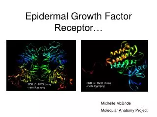
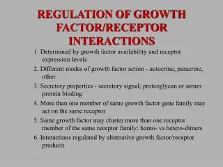
Sulindac metabolites inhibit epidermal growth factor receptor activation and expression. Heather A Pangburn*1,2, Hanna Kraus3, Dennis J Ahnen2,3,4 and Pamela L Rice2,3,4 Journal of Carcinogenesis 2005, 4 :16. Abstract.

E N D
Sulindac metabolites inhibit epidermal growth factor receptor activation and expression Heather A Pangburn*1,2, Hanna Kraus3, Dennis J Ahnen2,3,4 and Pamela L Rice2,3,4 Journal of Carcinogenesis 2005, 4:16
Abstract Nonsteroidal anti-inflammatory drugs(NSAIDs)可以用來降低colorectal cancaer (CRC)的死亡率,NSAIDs可以誘導CRC細胞進行apoptotic,並且可以抑制腫瘤細胞的生長。NSAIDs sulindac的代謝產物可以 downregulate extracellular-signal regulated kinase ½(ERK1/2) ,而造成CRC細胞進行apoptosis。我們這個實驗的主要目的是要來證實我們的假說, sulindac metabolites可以抑制或促進epidermal growth factor (EGF) receptor (EGFR)表現,因而影響到extracellular-signal regulated kinase ½(ERK1/2) 的表現,而達到抑制CRC細胞的生長的效果。 我們主要是利用HT29 human colon cancer cell來進行實驗,利用在細胞的培養基中加入EGF、 sulindac sulfide 或 sulindac sulfone來培養,並利用western immunoblotting 觀察細胞中的 phosphorylated EGFG、total EGFG、 phosphorylated ERK1/2、total ERK1/2、activated caspase-3和α-tubulin的表現量,和 nuclear morphology來觀察 apoptotic cells 和存活的細胞數量來判斷 sulindac對CRC細胞生長的影響情形。
Introduction 在美國CRC是最常見cancer第二死因,每年約有104950個案例,死亡率約56.29。在美國人的一生中平均約有6%的機會會得到CRC,所以作者想要研究NSAIDs這類藥物對於CRC的影響,看是否可以降低CRC的死亡率。 Sulindac在肝臟可以快速的代謝成兩種產物 sulindac sulfide 和 sulindac sulfone,我們將對此兩種藥物對於CRC 細胞的影響加以研究,其中sulindac sulfone並不屬於NSAIDs這類藥物,但他對於CRC 細胞的生長還是有影響,我們將再接下來的實驗中為大家證實。剛開始認為NSAIDs sulindac sulfide會抑制CRC細胞生長主要是因為他可以抑制cycloxygenase-1 (cox-1)或cox-2的活性,因而使細胞進行apoptosis但在我們後來的實驗中發現,最主要是因為使EGFR的表現量下降,才會使細胞進行apoptosis。所以我們設計此實驗來證實sulindac sulfide 和 sulindac sulfone會影響EGFR的表現量,進而使ERK1/2的表現量下降,而使細胞進行apoptosis。
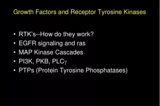
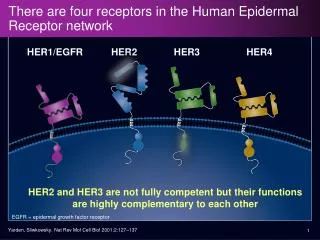
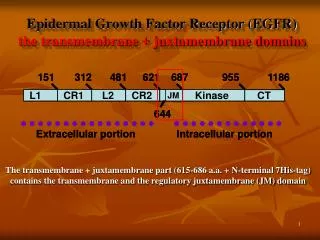
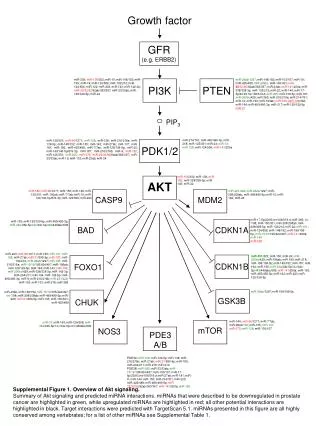
The EGFR/MAPK/ERK1/2 signaling pathway. We have previously shown that sulindac metabolites inhibit both MEK1/2 and ERK1/2 activation. The goal of this study was to determine if this inhibition is due to downregulation of the EGFR.
Material and method Morphologic quantitation of apoptotic cell death 利用 ethidium bromide/acridine orange double-dye morphological assay來觀察有多少細胞存活和多少細胞進行apoptosis,來做數量的統計。 western immunoblotting 1.將細胞從plates上取出,利用冰的phosphate buffer saline (PBS)洗一次。 2.2400g離心 5min去上清液,再重複上述步驟兩次,注意在所有的過程中都要盡量保持低溫。 3.在將離心所得的pellet放置到extraction buffer 30min on ice,每十分鐘取出來vortex一下。 4.離心18000g 4℃ 10min 取上清液,在利用Lowry的方法來测蛋白質濃度(約50μg)
5.利用sodium dodecyl sulfate-polyacrylamide gel electrophoresis(SDS PAGE)來分離蛋白質,再利用electrotansferred overnight onto immobilon-P polyvinylidene difluoride membranes上。 6.在將membranes浸泡在含有Tris-neutral saline with 1% dry milk and 0.05% Tween 20 30min來做 blooking。 7.取出並放入含有phosphorylated EGFG、total EGFG、 phosphorylated ERK1/2、total ERK1/2、activated caspase-3和α-tubulin primary antibody的溶液37℃下作用1小時 8.取出之後放到含有secondary antibody溶液中作用1小時。 9.放置含有 chemiluminescent substrate中作用一分鐘 10.在將所得到的結果拿去测光密度即可知道各種不同的蛋白之含量。
EGF induces EGFR and ERK1/2 phosphorylation. HT29 human colon cancer cells were grown to 80% confluence in medium containing 10% FBS then serum deprived for 48 h before treatment with vehicle (water), 10, or 100 ng/ml EGF. Cells were harvested 10 min after EGF treatment and lysates prepared for (A) Immunoblotting with antibodies raised against pEGFR(pY1068), total EGFR, ERK1/2, and total ERK1/2; α-tubulin immunoblots of the same lysates served as loading controls. Thegraphs show the densitometry results of the pEGFR bands (B) and pERK1/2 bands (C) normalized for the loading controls.
Dose response of sulindac sulfide inhibition of EGFR. HT29 cells were grown to 80% confluence in medium containing 10% FBS then serum deprived for 24 h before addition of drug. Cells were then treated with vehicle (0.1% DMSO), 40, 80, 120, or 160 μM sulindac sulfide, drug doses previously shown to induce apoptotic cell death in these cells. Twenty four hours after drug treatment, vehicle or 10 ng/ml EGF was added and cells were harvested 10 min later. (A) Immunoblots were performed on cell lysates with antibodies raised against pEGFR (pY1068), total EGFR, pERK1/2, total ERK1/2, and caspase-3; total ERK1/2 immunoblots served as loading controls. The graphs show the densitometry results of the pEGFR bands (B) and total EGFR bands (C). Results shown in figure are representative of 3 separate experiments.
Dose response of sulindac sulfone inhibition of EGFR. HT29 cells were grown to 80% confluence in medium containing 10% FBS then serum deprived for 24 h before addition of drug. Cells were then treated with vehicle (0.2% DMSO), 200, 400, 600, or 800 μM sulindac sulfone, drug doses previously shown to induce apoptotic cell death in these cells. Twenty four hours after drug treatment, vehicle or 10 ng/ml EGF was added and cells were harvested 10 min later. (A) Immunoblots were performed on cell lysates with antibodies raised against pEGFR (pY1068), total EGFR, pERK1/2, total ERK1/2, and caspase-3; total ERK1/2 immunoblots served as loading controls. The graphs show the densitometry results of the pEGFR bands (B) and total EGFR bands (C). Results shown in figure are representative of 3 separate experiments.
Dose response and time course of sulindac sulfide inhibition of EGFR. HT29 cells were grown to confluence in medium containing 10% FBS and treated with vehicle (0.1% DMSO), 160, or 180 μM sulindac sulfide for 1 h, 12 h, and 24 h. Cells were then harvested and immunoblots were performed on cell lysates with antibodies raised against pEGFR (pY1068), total EGFR, pERK1/2, total ERK1/2, and cleaved caspase-3; α-tubulin immunoblots of the same lysates served as loading controls.(A) 1 h, 12 h, and 24 h immunoblot results. The graphs show the densitometry results of the pEGFR bands (B) and total EGFR bands (C).
Dose response and time course of sulindac sulfone inhibition of EGFR. HT29 cells were grown to confluence in medium containing 10% FBS followed by treatment with vehicle (0.2% DMSO), 400, or 600 μM sulindac sulfone for 1 h, 12 h, and 24 h. Cells were then harvested and immunoblots were performed on cell lysates with antibodies raised against pEGFR (pY1068), total EGFR, pERK1/2, total ERK1/2, and cleaved caspase-3; α-tubulin immunoblots of the same lysates served as loading controls. (A) 1 h, 12 h, and 24 h Western blot results. The graphs show the densitometry results of the pEGFR bands (B) and total EGFR bands (C).
Effect of the caspase inhibitor, ZVAD, on apoptosis and inhibition of EGFR. HT29 colon cancer cells were grown to confluence in medium containing 10% FBS followed by pretreatment with or without 25 μM zvad for 1 h. Cells were then treated with vehicle (0.2% DMSO) or 600 μM sulfone for 48 h. Cells were harvested and immunoblots were performed on cell lysates with antibodies raised against pEGFR (pY1068), total EGFR, and cleaved caspase 3; α-tubulin immunoblots of the same lysates served as loading controls. The graphs show morphological apoptosis results (A) 48 h Western immunoblot results (B) and densitometry of the pEGFR bands (C) and total EGFR bands (D).
Result 1.sulindac sulfide 和 sulindac sulfone都可以使EGFR的活性降低,而使CRC細胞進行apoptosis,但sulindac sulfone的效果比較沒有這麼好。 2.EGFR被抑制並不是因為caspase-3被活化的原因。 3.sulindac是直接使EGFR的表現量下降。而跟COX沒有關係。