Download
1 / 33
430 likes | 1.4k Views
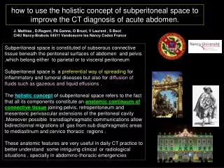
how to use the holistic concept of subperitoneal space to improve the CT diagnosis of acute abdomen. . J. Mathias , D.Regent, PA Ganne, O Bruot, V Laurent , S Beot CHU Nancy-Brabois 54511 Vandoeuvre les Nancy Cedex France .

E N D
how to use the holistic concept of subperitoneal space to improve the CT diagnosis of acute abdomen. J. Mathias , D.Regent, PA Ganne, O Bruot, V Laurent , S Beot CHU Nancy-Brabois 54511 Vandoeuvre les Nancy Cedex France Subperitoneal space is constituted of subserous connective tissue beneath the peritoneal surfaces of abdomen and pelvis ,which belong either to parietal or to visceral peritoneum Subperitoneal space is a preferential way of spreading for inflammatory and tumoral diseases but also for diffusion of fluids such as gazeous and liquid effusions . The holistic concept of subperitoneal space refers to the fact that all its components constitute an anatomic continuum of connective tissue joining pelvis, retroperitoneum and mesenteric perivascular extensions of the peritoneal cavity .Moreover possible transdiaphragmatic communications allow bidirectionnal migrations of gas from sub diaphragmatic areas to mediastinum and cervico thoracic regions . These anatomic features are very useful in daily CT practice to better understand some intriguing clinical or radiological situations , specially in abdomino-thoracic emergencies
subperitoneal anterolateral or properitoneal space subperitoneal perivascular mesenteric space subperitoneal anterolateral or properitoneal space subperitoneal anterolateral or properitoneal space fascia transversalis 1.anatomical basis of subperitoneal space anterior abdominal wall ,dorsal view retroperitoneal compartments retropubic (Retzius) subperitoneal space
posterior mediastinal connective tissue anterior extrapleural space properitoneal space retroperitoneal space 3 compartments: -anterior pararenal -périrenal -posterior para renal retropubic subperitoneal space (Retzius' space) there is a continuum between subperitoneal connective tissue of anterior abdominal wall, pelvis and retroperitoneum (behind posterior parietal peritoneum).. In the thorax, extra pleural space and mediastinum are also made of connective tissue with potential communications (oesophageal and aortic hiatus. between diaphragmatic anterior insertions) pelvic subperitoneal space
If connective tissue of subdiaphragmatic abdomen (peritoneal cavity and retroperitoneum )is extracted , the continuum is demonstrated . We have to consider that subperitoneal space extends along mesenteric vessels in the mesentery and the mesosigmoid ( ) ,coeliac trunk and coronary ligament ( ) possible direct or inverse migration of gas from subperitoneal space to subpleural or mediastinal connective tissue
tuberculous ascitis in a 43 yo african man • continuum between: • -subperitoneal perivascular space (mesentery mesosigmoid ) • -retroperitoneal space • subperitoneal pelvic space • properitoneal space
two cases of cirrhotic ascitis to depict the potential road for gaseous diffusion from subperitoneal perivascular space of the mesosigmoid to its mesenteric counterpart (yellow road) or to posterior mediastinal connective tissue (green road) ,through aortic and/or oesophageal foramens properitoneal space subperitoneal mesosigmoid space great omentum floating in the supra mesocolic area cirrhotic ascitis in a 57 yo man cirrhotic ascitisin in a 48 yo man
2. fluid diffusion through connective subserous spaces of the trunk 2a. gazeous migrations in subserous spaces of the trunk they are the most important and fortunately the easiest images to see and to analyse on CT of acutely ill patients but they can be confusing if they are misdiagnosed. The first step is tolearn how to recognize the exact anatomic situation of these gazeous images • how can we identify pro peritoneal gazeous images ? the easiest way to avoid misinterpretation of pro peritoneal gazeous images is to consider situations in which nothing has been done in the abdomen ! -massive subcutaneous thoracic emphysema -typical images of properitoneal gas ( ) (symetrical linear collections of gas sparing the median region) -retropneumoperitoneum ( ) -no pneumoperitoneum ! massive subcutaneous cervico thoracic emphysema after surgical lung biopsy
-pneumothorax ( ), pneumopericardium ( ) and subcutaneous anterior thoraco-abdominal emphysema ( ) • properitoneal gas is seen ,lining the anterior edge of the liver , thus mimicking pneumoperitoneum ( ) ,however these images are fixed in their position and are not situated at the apex of the peritoneal cavity as in pneumoperitoneum . • -pneumothorax in the anterior costo-diaphragmatic recess ( ) lies before the diaphragm and also need to be differenciated from pneumoperitoneum massive subcutaneous emphysema of the antero superior thoracic wall and the cervic thoracic junction after a difficult transjugular implantation of à Port-a Cath.
how can we identify intra peritoneal gazeous images ? -identifying a pneumoperitoneum on CT scan is generally very easy specially with large peritoneal gazeous effusions (ulcer perforation, caecal diastatic perforation, bowel wall endoscopic perforation…) are present . - when you are in doubt (specially with small gazeous collection of the subphrenic space ) ,remember that the only sign whicht will never fail you is the outlining of the falciform ligament by free intra peritoneal gas ( ) ! ! ! anterior gastric wall perforated ulcer
-when confronted with juxta abdominal wall gazeous images , you hesitate between pro or intra peritoneal gas ( ) -look at the highest point of the peritoneal cavity which is always located in the median zone and you will see gas bubbles lining anterior parietal peritoneum ( ) in case of true pneumoperitoneum perforated anterior duodenal ulcer ; alcoholic cirrhosis with ascitis , in a 62 yo woman
juxta splenic gazeous images ( ) belong to retro-pneumoperitoneum as they stay between splenic capsule and visceral splenic peritoneum • how can we identify retro peritoneal gazeous images ? -propagation of gas from cellulitis through subperitoneal pelvic space ( ) to retroperitoneum ( ) -no free gas is observed in the peritoneal cavity -gazeous retropancreatic and juxta splenic images ( ) belong to retroperitoneal space ! ! ! rheumatoid arthritis ,diabetic , coticotherapy ,sepsis of the ischiatic region (cellulitis after skin disruption),i, a 58 yo man
-iatrogenic retro-pneumoperitoneum secondary to instrumental rupture of the posterior duodenal wall ,at the level of the major duodenal papilla ( ) (extra hepatic biliary tree has been opacified before sphincterotomy ). -the interesting fact is the presence of gas in antero lateral properitonealspace ( ) and Retzius space ( ). -no pneumoperitoneum is observed despite the volume of gas visible on these images ! ! ! and this situation may theoricallybe managed without surgery…. acute abdomen after endoscopic extraction of a choledcolithiasis in a 66 yo woman .
don't be afraid when you see a lot of gas on CT of acute abdomen and make a thorough analysis! ! -posterior pneumomediastinum ( ) and retro-pneumoperitoneum ( ) due to lower oesophageal mechanical rupture after a failed attempt to extract a piece of impacted meat ( ). -gas migrated to posterior mediastinum via peri oesophageal hiatus ( ) ; -no gas migrated around IVC due to a circular continuous fibrous membrane joining venous wall and diaphragm that prevent transdiaphragmatic road at this point . -perivisceral gas bubles correspond to migration in subperitoneal perivascular spaces ( ) painful aphagia after a failed attempt to extract an impacted piece of meat in the lower oesophagus
fever, subcutaneous emphysema of the right axilla in a 71 yo man (1) -CT confirm right axilla subcutaneous emphysema ( red circle ) and pneumomediastinum with total homogeneous gaseous dissection of mediastinal connective tissue ,from top to bottom ( ) -we can see gas around the IVC ( ) and in the retroperitoneum ( ) -in front of the liver it is diificult to determine the exact location of the gaseous images ( ) (anterior pleural recess? pro peritoneal gas ?pneumoperitoneum?)
fever, subcutaneous emphysema of the right axilla in a 71 yo man (2) -the retro-pneumoperitoneum is obvious ( ) with gas around caudal pancreas and sagittal reformation shows that this gas is coming from mesosigmoid ( ) following a ruptured sigmoid diverticule -a subsidiary question concern a large amount of gas behind anterior abdominal wall ( ) .Laparoscopy confirm a pneumo-omentum with gazeous infiltration of epiploic and pericolic appendages.
59 yo man ,returning from an endoscopic resection of recto-colic polys ;a nurse of the post anesthesia care unit is puzzled by an aspect of the penis she is not familiar with…; clinical examination reveals subcutaneous cervuco-thoracic emphysema (1) • -CT confirms subcutaneous cervical emphysema, ( ), and pneumomediastinum ( ) • extensive retro-pneumoperitoneum ( ) , • -pneumopenis ( ) and gazeous dissection of groin subcutaneous connective tissue ( )
this very impressive "total body" gazeous dissection of retroperitoneum ( ) and mediastinum ( ) after a common endoscopic resection of a pediculated recto-sigmoid polyp give us a good illustration of the holistic concept of subperitoneal space an its relations with mediastinal connective tissue . Note the gazeous dissection of the fascia of Treitz with "floatting" duodeno pancreas ( orange circle ). 59 yo man ,returning from an endoscopic resection of recto-colic polys ;a nurse of the post anesthesia care unit is puzzled by an aspect of the penis she is not familiar with…; clinical examination reveals subcutaneous cervuco-thoracic emphysema (1)
endoscopic sphincterotomy and transpapillary extraction of biliary stones of the main bile duct in a 72 yo woman ; incresasing pain with abdominal guarding -CT confirms the retro-pneumoperitoneum ( ) due to posterior rupture of the juxta papillary duodenal wall. -but dont forget to look for the sign of associated pneumoperitoneum : free pre hepatic gas outlining the falciform ligament ( ) which indicate surgical treatment .
pain and tenderness of the right iliac fossa; ; moderate fever in a 80 yo man -no pneumoperitoneum! ! -retroperitoneal abcess ( ) due to retrocaecal appendicitis ( ) -gazeous diffusion in properitoneal space ( ) and in subperitoneal space of the diaphragm ( ) courtesy of M Lamrani MD Les Sables d'Olonne
peri rectal abcess operated 3 days ago in a 69 yo man ; recurrent fever and persistant local sepsis -be careful ! there is no pneumoperitoneum -all visible gaz bubbles belong to retropneumoperitoneum ( ) or to mesenteric subperitoneal space ( ) -small pneumomediastinum ( ) -peri rectal abcess ( )
respiratory distress during a tough endoscopic extraction ot a main bile duct stone in a young 39 yo woman ; subcutaneous cercico-thoracic emphysema -retropneumoperitoneum ( ) due to traumatic tear of the juxta papillary postérior duodenal wall -gas in the properitoneal space ( ) and in subperitoneal pelvic space and deep perineal connective tissue ( ) -bilateral large gazeous subpleural effusion ( ) -no pneumoperitoneumand no pneumothorax are noticed ! ! !
young girl with psychological problems , who auto-injected syringes of air in her back ( ); you might also suggest that she is probably right-handed! ! ! ! young teenager , coming to emergency room for giant subcutaneous emphysema without any other complaint CT confirms X ray plain films : huge extensive retropneumoperitoneum ( ) a global and harmonious pneumomediastinum ( ) and subcutaneous emphysema ( ) can you guess the origin with this last picture
59 yo man, heavy smocker , ,laryngeal cancer two years ago, intermittent abdominal pain. COPD and severe emphysema -cystic chronic pneumatosis coli is a condition very often observed in association with chronic hypoxemia , specially heavy smokers with COPD . Excess pas is coming from bowel lumen ,due to modified bacterial population secondary to chronic hypoxemia and insufficient methane degradation. -cystic gaseous lesions are generally developed in submucosa but ,as in this case, also in subserous connective tissue ( );. -a retropneumoperitoneum ( ) is very frequently associated and always in the right side. It corresponds to migration of gas from parietal lesions through subperitoneal space of right Toldt's fascia or of the right transverse mesocolon . -pneumoperitoneum ( ) secondary to spontaneous rupture of subserosal gazeous cyst is also frequent
chronic mild abdominal pain , heavy smoker ; severe hypoxemia at arterial blood gas sampling in a 52 yo woman -typical chronic cystic pneumatosis coli linked to chronic hypoxemia ( ). -massive gazeous dissection of the right Toldt's fascia ( ) -right retropneumoperitoneum ( ) with diffusion of gas bubbles up to diaphragmatic subperitoneal space ( ). courtesy of A Rodde MD Luxembourg
53 yo man , heavy smoker with mild abdominal pain , operated after discovery of a pneumoperitoneum on CT ; no intestinal perforation at surgery ! -typical case of chronic cystic pneumatosis ( ) of the small bowel due to chronic anoxemia ( COPD ) -small bowel chronic cystic pneumatosis is preferentially located in subserosis .Thus it represents the main cause of "medical" or "non surgical" pneumoperitoneum ( ) -large retropneumoperitoneum is an excellent diagnostic argument ( ) -remember:"don't operate images ! ! ! " courtesy of M. Deneuville MD Metz
58 yo man with chronic respiratory insufficiency due to major biapical tuberculous sequellae . • asymptomatic pneumoperitoneum ( ) and retropneumoperitoneum ( ) • small cluster of jejunal chronic cystic subserous pneumatosis ( ) at the origin of gazeous migration • subtle details but big diagnostic impact ! ! !
2a. liquid migrations through subperitoneal space of the trunk Instances of loco-regional liquid fluiddiffusion are more commonly seen,, for exemple in acute exsudative pancreatitis and in diffusion of retro peritoneal or intra peritoneal infectious processes. -diffusion of peripancreatic fluid ( ) through subperitoneal perivascular space produces a real hydrodissection of the mesentery ( ) , up to the distal segment of the superior mesenteric artery and right Toldt's fascia ( ).. -para renal anterior compartment of the retroperitoneal space is also involed ( ) acute pancreatitis of unknown origin , without pancreatic necrosis in a 34 yo man
54 yo alcoholic man with , acute pancreatitis and spontaneous hyperdense pseudocyst ( ); CT confirms a pseudo aneurysm of a pancreatico-duodenal artery ( ). peripancreatic migration of fluid follows subpleural space of ligaments and mesos to reach : -right transverse mesocolon via duodeno colic ligament ( ) -right Toldt's fascia and ascending colon ( ) -pars pediculosa of gastro hepatic ligament (lesser omentum) ( )
2. inflammatory and tissular spreading through connective subserous spaces of the trunk considerable work has been done to show the importance of subserosal space to understand the spread of tumoral and inflammatory diseases in abdomino-pelvic cavity. We would like to show 2 pertinent didactic examples 64 yo man ,heavy smoker with weight loss , postprandial epigastric pain and vomiting ;clinically suspected of acutizing chronic mesenteric ischemia -high grade stenosis of the third portion of the duodenum ( ) -coexisting with thick mural thrombus of an atheromatous abdominal aorta ( ) -no main pancreatic duct dilatation was observed ( ) . -next slide will show more detailed images
-we can see a typical aspect of fibrous periarterial extension of a pancreatic ductal adenocarcinoma ( ) -the pancreatic lesion has developed from the uncus and spread down along the mesenteric perivascular subperitoneal space ( ) -dilatation of mesenteric distal veins upstream fibrotic meseteric expansions are highlighted on MIP coronal and axial images ( ).
56 yo man ,misdiagnosed and treated for 16 years for tuberculosis , with liver and spine( L1-L2 )locations. • diagnosis of echinococcosis multilocularis was finally established after surgery for medullary compression • initial calcified lesion of the left liver ( ) migrated through the subpleural space of the lesser omentum to retroperitoneum (para renal anterior and para renal posterior compartments ) ( ) • -this parasitic fibrous aggressive reaction extended to lumbar spine determining the "pseudo Pott" aspect ( )
3 conclusions and take home messages - becaming more familiar with the holistic concept ofsubperitoneal space is very helpful to read CT images of acute abdomen , particularly for exact determination of the localisation of gazeous images . -it is often necessary to to have readily available CT thoraco -abdomino-pelvic examination in order to avoid heavy consequences of misinterpration in fluid migrations (specially gazeous ) in subperitoneal space -don't forget to throughly analyse and identify gazeous images of properitoneal space and don't misinterpret them as pneumoperitoneum -always look for retropneumoperitoneum when you have problems with gazeous bubbles anywhere in the trunk ; it is a very important item to take into account for correct diagnosis -subperitoneal space is an Ariane's thread that helps the radiologist to recognize fluid migration roads through the labyrinth of abdomino thoracic anatomy -thank you for showing interest in this poster holistic concept of subperitoneal space in yellow ! from ref 8.Pickhardt PJ , Bhalla S Usual nonneoplastic peritoneal and subperitoneal conditions : CT findings RadioGraphics 2005 ;25 : 719-730
References 1.Oliphant M , Berne AS , Meyers MA Imaging the direct bidirectionnal spread of disease between the abdomen and the female pelvis via the subperitoneal space Gastrointest Radiol 1988 ;13 , 285-288 2.Oliphant M , Berne AS , Meyers MA Bidirectionnal spread of disease via the subperitoneal space .The lower abdomen and left pelvis Abdom. Imaging 1993;18:117-125 3.Oliphant M , Berne AS , Meyers MA Spread of disease via the subperitoneal space Abdom. Imaging 1993 ; 18 , 109-116 4.Oliphant M , Berne AS , Meyers MA Direct spread of subperitoneal disease inti solid organs Abdom. Imaging 1993 ; 20 : 141 -147 5.Oliphant M , Berne AS , Meyers MA The subperitoneal space of the abdomen and pelvis Amer J Roentgenol AJR 1996 ; 167 :1433-1439 6.Silverman PM The subperitoneal space: mechanism of tumor spread in the peritoneal cavity,mesentery and omentum Cancer Imaging 2003 ; 4 : 25-29 7.Oliphant M , Berne SA , Meyers MA The subperitoneal space:; normal and pathologic anatomy in Meyers MA Dynamic radiology of the abdomen 5th edition 2000 Springer ed.,607-634 8.Pickhardt PJ , Bhalla S Usual nonneoplastic peritoneal and subperitoneal conditions : CT findings RadioGraphics 2005 ;25 : 719-730