Download
1 / 1
10 likes | 165 Views
BACKGROUND. Microfabricated bioreactors offer significant advantages over traditional cell culture techniques: constrained μ-scale dimensions small volumes and cell numbers physiologically relevant environment Our VIIBRE developed bioreactors are:

E N D
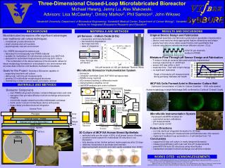
BACKGROUND • Microfabricated bioreactors offer significant advantages over traditional cell culture techniques: • constrained μ-scale dimensions • small volumes and cell numbers • physiologically relevant environment Our VIIBRE developed bioreactors are: • fabricated from biologically inert PDMS material • suitable for multicelluar 3D cell structure cultures (e.g. MCF10A) • designed for automated measurements and long-term culture • The combination of the above features of the bioreactor allows for tissue morphology formations in a simulated in vivo environment with accurate data collection and feedback-mediated homeostasis. • Goals for this Project- Develop a Bioreactor capable of: • supporting long-term cell culture • taking near real time pH measurements • growing 3D multicellular morphological structures • maintaining constant nutrient supply and waste removal y = -70.647x + 680.15 R2 = 0.999 Flow-Through IrOx pH Sensor pH Acidified Media Bioreactor Iridium Oxide pH-Sensing Electrode Faraday Cages Three-Dimensional Closed-Loop Microfabricated Bioreactor Michael Hwang, Jenny Lu, Alex Makowski, Advisors: Lisa McCawley1, Dmitry Markov2, Phil Samson2, John Wikswo Vanderbilt University, Department of Biomedical Engineering; Vanderbilt Medical Center, Department of Cancer Biology1; Vanderbilt Institute for Integrated Biosystems Research and Education2 + IA Iridium Oxide Quasi-Reference Electrode - Bias Current Return Path DAQ Device Cavro XLP 6000 Syringe Pumps Cell Media Reservoir pH 6 Calibration Solution pH 8 Calibration Solution 1.875 in. 47mm centered 8mm centered MATERIALS AND METHODS RESULTS AND DISCUSSIONS Original Device Design and Fabrication • generated assembly and fabrication protocols repeatable characteristics • device cross-sections show appropriate feature dimensions and shape • gravity feed tests show viable sealed fluidics and quantifiable flow • volume calculations indicate maximum diffusion volume <12 μL pH Sensors – Iridium Oxide (IrOx) • inexpensive and manufacturable • low sensitivity degradation • fast response time • ease of integration 100x100 μm channels molded in PDMS Potential (mV) Miniature Flow-Through pH Sensor Design and Fabrication • Ti-Iridium Oxide ph sensors fabricated and fully characterized • average responsivity of >60mV/pH • sensor drift was within ± 2mV • response time ~7sec with 2.0pH change • no significant hysteresis IrOx pH sensors on 125 μm diameter Titanium Wires Potential (mV) Microfluidic Bioreactor Instrumentation System • bioreactor • computer controlled Cavro XLP 6000 syringe pumps • PDMS mixing manifold • IrOx sensors working in differential mode • instrumentation amplifier circuit • LabVIEW analysis and control software Graph of bioreactor pH measurements during pumping indicates cell health Time (sec) MCF10A Cells Formed Acini in Bioreactor Culture Well Optimized Concentration of Cells for Culture Chamber: ~1500 cells seeded MATERIALS AND METHODS Bioreactor Components • inert PDMS cell growth chamber contains Matrigel matrix and cells • nanopore filter provides diffusive nutrient exchange and prevents clogging • microfluidic supply network provides media/waste exchange • fluidic ports in acrylic lid interfaces device with pumps • brass clamp provides structural integration Acinar morphology in bright field image (left) confirmed by Confocal “Z-stack” (right) Access Ports Microfluidic Instrumentation System Developed LabVIEW modules for: • automated sensor calibrations • pH measurements • Cavro pump control Plexiglas 3mm 2mm Channel Layer - PDMS 300μm Matrigel with Cells (8mm diam.) 1mm Glass Slide ~1.9 in. square Future Directions • run fully sterilized integrated bioreactor for 20 Days • optimize low volume pH measurements and fabricate tube IrOx sensors • implement feedback loop automation (<.4 pH window from 7.0) 3D Culture of MCF10A Human Breast Epithelials • epithelial cells are the origin of 80% of all breast tumors (Knauss) • excellent model system for multicellular 3D structure in our bioreactor • fully develop acinar (hollow sphere) mammospheres after 20 days • relatively inexpensive to purchase and maintain • Approved growth protocols and cells readily available from ViCBC Conclusions • supported 20 day cell culture in newly designed bioreactor • measured acidification with near real time pH measurements • grew MCF10A acini 3D structures using microfluidics • maintained consistent nutrient exchange via LabView modules WORKS CITED / ACKNOWLEDGEMENTS Debnath, J., Brugge, J. S., Modeling glandular epithelial cancers in three-dimensional cultures, Nature Reviews Cancer 5, 675-688 (2005). Knauss, U. “Cell Growth Control of Breast Epithelial Cells”. Abstract. California Breast Cancer Research Program. Differential expression and subcellular and localization of the GTPase Rac3. FASEB Meeting: Small G-Proteins & Cell Dynamics, 2002. Special Thanks to: Ron Reiserer, Eduardo Lima, Igor Ges, Franz Baudenbacher, Don Berry, S. Marzouk, David Schaffer, David Shifrin, Bryan Gorman, Steven Manuel for device machining, and Dr. King and NCIIA for additional funding. Illustration of Acinar Morphology Formation (from Debnath and Brugge 2005)