The Heartbeat
The Heartbeat. Heartbeat. Each heartbeat is called a cardiac cycle. First the two atria contract at the same time. Next the two ventricles contract at the same time. Then all the chambers relax. Systole – contraction of the heart muscle Diastole – relaxation of the heart muscle.


The Heartbeat
E N D
Presentation Transcript
Heartbeat • Each heartbeat is called a cardiac cycle. • First the two atria contract at the same time. • Next the two ventricles contract at the same time. • Then all the chambers relax. • Systole – contraction of the heart muscle • Diastole – relaxation of the heart muscle.
Each heartbeat last about 0.85 seconds so the average person’s heart beats about 70 times a minute.
The Sound of a Beating Heart • When the heart beats it makes the familiar “lub-dup” sound. • The “lub” is long and low pitched. This sound is caused by the atrioventricular valves (bicuspid and tricuspid) closing as the ventricles contract. • The “dup” is shorter and sharper. This sound is caused by the semilunar valves (pulmonary and aortic) closing after the blood is pumped out of the ventricles into either the pulmonary artery or the aorta.
Intrinsic Control of the Heartbeat • The rhythmical contraction of the atria and ventricles is due a conducting system inside the heart. • The conducting system is made up of the following parts:
1) two nodes (small regions of neuromuscular tissue) • SA (sinoatrial) node – located in the upper, dorsal wall of the right atrium. Initiates the heartbeat (also known as the pacemaker) • AV (atrioventricular) node– located at the base of the right atrium. Transmits impulses received from the SA node to the ventricular walls.
2) Conducting bundle- specialized fibres that conduct impulses from the AV node to the ventricles. The conducting bundle is also known as the Bundle of His. • Divide to form the right and left bundle branches which extend into the right and left ventricles. • The terminal branches of the bundle braches are called Purkinje fibers.
The SA node initiates the heartbeat and automatically sends out an excitation impulse every 0.85 seconds which causes the atria to contract. • When the impulses reach the AV node there is a slight delay as the impulse passes through the bundle of His that allows the atria to finish their contraction before the ventricles begin to contract.
The signal for the ventricles to contract travels from the AV node through the branches of the bundle of His to the Purkinje fibers. This pathway efficiently causes the ventricles to contract. • The SA node is called the pacemaker because it usually keeps the heartbeat regular.
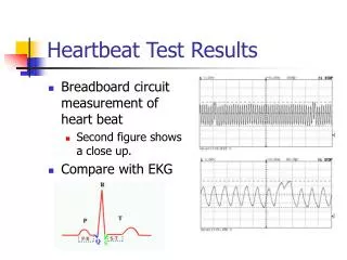
Electrocardiograms (ECG) • A recording of the electrical changes that occur in the myocardium during a cardiac cycle. • Electrodes are placed on the skin and are connected by wires to an instrument that detects and records the myocardium’s electrical changes. • Electrical changes in the myocardium can be detected on the skin’s surface because body fluids contain ions that conduct electrical currents from the heart throughout the body.
ECG • A normal ECG contains three distinct sections: • P wave – atrial fibers produce an electrical change. This indicates that the atria are about to contract. • QRS complex – signals that the ventricles are about to contract. • T wave – occurs when the ventricles are recovering from contraction.
The above diagram of an irregular ECG displays a condition known as ventricular fibrillation. • This condition causes uncoordinated contraction of the ventricles. • This condition can be caused by an injury or drug overdose. • Once the ventricles are fibrillating, they have to be defibrillated by applying a strong electrical current for a short period of time. • This current allows the SA node to reestablish a coordinated beat.
Once the ventricles are fibrillating, they have to be defibrillated by applying a strong electrical current for a short period of time. This current allows the SA node to reestablish a coordinated beat.