Download

1 / 9
90 likes | 185 Views
ATOMIC FORCE MICROSCOPY OF HUNTINGTIN AGGREGATES. Tomas T Ding and Peter T Lansbury Jr Center for Neurologic Diseases, Brigham and Women’s Hospital 65 Landsdowne street, Cambridge, MA 02139. Jin Wang and James F Gusella Molecular Genetics Unit, Massachusetts General Hospital

E N D
ATOMIC FORCE MICROSCOPY OF HUNTINGTIN AGGREGATES Tomas T Ding and Peter T Lansbury Jr Center for Neurologic Diseases, Brigham and Women’s Hospital 65 Landsdowne street, Cambridge, MA 02139 Jin Wang and James F Gusella Molecular Genetics Unit, Massachusetts General Hospital 149 13Th Street, Charlestown, MA 02129
ABSTRACT • Background: The extent of in vitro formation of aggregate with amyloid characteristics by the N- terminus of huntingtin with glutamine repeat lengths in the pathological range correlates inversely with the age of onset of Huntington’s disease. Furthermore, amyloid has been detected in HD-brain inclusions . Finally, huntingtin aggregation, like aggregation of other amyloidogenic and disease-related proteins such as amyloid-b peptide and a-synuclein, may follow a nucleation-dependent polymerization pathway. Because both amyloid-b peptide and a-synuclein form pre-fibrillar and possibly neurotoxic aggregates in vitro we speculate that mutant huntingtin assembles into protofibrils and that those could play a role in the onset of Huntington’s disease. • Methods: We utilize atomic force microscopy (AFM) to image huntingtin aggregates adsorbed to mica. In AFM a substrate is scanned with a sharp tip attached to a cantilever while the deflections of the cantilever are translated into a ‘relief-map” of the surface. Such maps provide us with 3-dimensional morphological information about single proteins and protein aggregates. • Results and conclusions: Huntingtin fibrils feature a diameter of 7-10 nm, similar to the fibrils described in the context of Alzheimer’s and Parkinson’s disease, regardless of glutamine repeat length. Preliminary data implies that a higher number of fibrils (eg per unit volume) is formed whereas the lengths of the fibrils remain relatively constant on increasing glutamine repeat number; suggesting that the nucleation rate increases with increased glutamine repeat length. We also have indications that aggregating huntingtin forms pre-fibrillar aggregates in vitro. Thus, within 2h Q41 huntingtin produces spherical assemblies with an average height of 2.4 nm, similar to the “spheres” detected in aggregating a-synuclein . Apparently, this distribution is not stable but changes over time, so after 98h the average height increased to 3.4 nm as the measured particle density dropped from 400 to 20 particles per square micron. Since huntingtin Q41 is capable of forming both fibrils and a protofibril-like intermediate in vitro we argue that other huntingtin mutants also form such intermediates, and that they could be considered as possible drug targets.
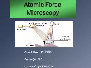
Atomic force microscopy (AFM) Examples: Carbon atoms Creates a relief map of the surface. Higher features are represented with brighter colors. b-Amyloid fibril
Red cursors (horizontal):131 nm Green cursors (height): 8.1 nm b-Amyloid fibril Individual Particles: Cross-section Histogram Populations: Mutant (A53T) a-Synuclein (protofibrils)
Q41 2h Q41 98h Huntingtin exon 1 incubated at 37°. The particles were counted and heights measured. n:2526, coverage 8.6% particle height: 3±2 nm n: 2342, coverage: 33% particle height: 5±3 nm
“Preliminary conclusion”: Particle-size distribution changes over time 2h Q41 98h Height distribution histograms taken at 2h and 98h. Note that this is relative amounts; the total particle number is the same for the two distributions.
Annular Q41 protofibrils? Cursors: 18 nm. Height 2.5 nm. Mean width 25 nm. Volume (10-18 cm3). Calculated weight corresponds to 56 Q41 monomers.
For comparison: a-Synuclein Protofibrils. “Spherical” (left) and Annular protofibrils formed by the a-synuclein A53T Mutant. This protein is Associated with familial Parkinson’s disease. The A30P mutant and wild type a-synuclein also form annular structures. Annular A53T (from AFM): Diameter 10-12 nm Height 1.9-2.3 nm. 100nm square 2.5m square Height: 3±1nm N: 2993 EM by Hilal A. Lashuel, Harvard Medical School. Scale bar 100 nm.
Summary Aggregating huntingtin rapidly forms spherical aggregates with a size distribution that changes over time. Preliminary data suggest that Q41 is Capable of forming annular “toxin-like” Structures similar to those formed by a-synuclein (and b-amyloid).