Download

1 / 76
770 likes | 1.2k Views
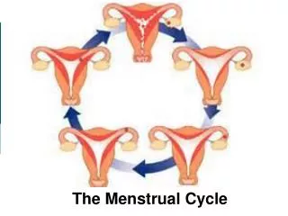
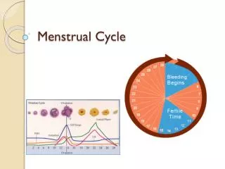
Higher level- Menstrual Cycle. Hormonal control in the menstrual cycle. Four hormones involved: FSH (Follicle Stimulating Hormone) Oestrogen LH (Luteinising Hormone) Progesterone Each hormone causes the production of the hormone following it and inhibits the hormone preceding it.

E N D
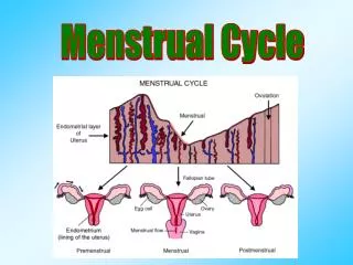
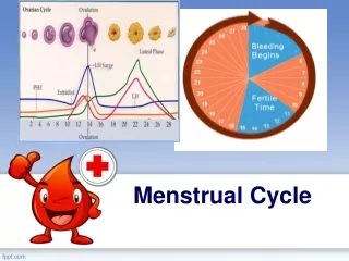
Hormonal control in the menstrual cycle • Four hormones involved: • FSH (Follicle Stimulating Hormone) • Oestrogen • LH (Luteinising Hormone) • Progesterone • Each hormone causes the production of the hormone following it and inhibits the hormone preceding it
FSH – Follicle Stimulating Hormone • Produced by pituitary gland • Produced early in the cycle (days 1-5) • Stimulates a few potential eggs to develop, surrounded by graafian follicles • Only one usually survives • Sometimes used in fertility treatments to stimulate ovaries to produce eggs – often lots of eggs develop. This explains some multiple births • Each graafian folllicle then produces oestrogen
Oestrogen • Produced by the graafian follicle in the ovary • Produced from days 5 -14 • Causes the endometrium to develop • Inhibits FSH ensuring no further eggs develop (useful in contraceptive pill) • High levels of oestrogen just before day 14 stimulate release of LH
LH - Luteinising Hormone • Produced by the pituitary gland • Produced on day 14 • Causes ovulation • Causes the remains of graafian follicle to develop into corpus luteum • Corpus luteum makes final hormone in the cycle progesterone (along with small amounts of oestrogen)
Progesterone • Produced by the Corpus Luteum in the ovary • Produced from days 14-28 • Maintains structure of endometrium • Inhibits FSH to stop further eggs developing • Inhibits LH to stop further ovulation and pregnancies • Prevents contractions of the uterus
Insemination • Insemination is the release of sperm into the female • Contractions of uterus and fallopian tubes move the sperm to the fallopian tubes within 5 minutes • If an egg is present it releases chemicals to attract the sperm this is called chemotaxis
Fertilisation Fertilisation is the fusion of the egg and sperm nuclei to form a diploid zygote. This usually occurs in the fallopian tube.
Fertilisation The acrosome releases enzymes to digest the egg membrane A number of sperm may reach the egg at the same time. The sperm loses its tail and the head enters the egg. The sperm and egg nuclei fuse to form a zygote A chemical reaction at the membrane prevents other sperm cells entering.
Implantation Implantation is the embedding of the fertilised egg into the lining of the uterus • This occurs 6 - 9 days after fertilisation. • By this time the zygote has grown into an embryo. • During implantation a membrane called the amnion develops around the embryo. This secretes amniotic fluid which will surround the developing embryo and act as a shock absorber. • After implantation the placenta forms.
Sequence of development from fertilised egg Early stages
Sequence of development from fertilised egg • The zygote contains 46 chromosomes, twenty three from the egg and 23 from the sperm
It divides rapidly by mitosis to produce 2 cells, then 4, then 8, 16 etc. and continues to divide
At this point the developing individual is referred to as the morula
Around 5 days after fertilisation the morula forms a hollow ball of cells called the blastocyst
The outer layer of the blastocyst forms the trophoblast. This will later develop into the layer of membranes that surround the embryo (placenta and amnion) Trophoblast
The inner cells (called the inner cell mass) of the blastocyst will eventually form the embryo. These cells are not yet specialised. They have a phenomenal ability to differentiate – divide to give rise to many different types of tissue Inner cell mass
The morula/blastocyst is pushed along the fallopian tube until it enters the uterus
Here it will implant into the uterus wall. The endometrium now provides nourishment for the developing blastocyst • Connections with the mother will begin to form (placenta and umbilical cord)
Sequence of development from fertilised egg Development of the embryo
About 10 days after fertilisation the inner cell mass forms the embryonic disc • This usually consists of three layers called germ layers • Ectoderm (outside) • Mesoderm (middle) • Endoderm (inside)
Each of these layers gives rise to specific structures in the developing embryo • In humans the mesoderm is split by a layer called the Coelom • This allows space for more complex organs such as heart, lungs and kidneys to develop
Ectoderm – skin, nervous system Coelom – heart, lungs Mesoderm – muscles, skeleton Endoderm – inner lining of digestive system
The Amnion • When first formed the amnion is in contact with the embryo, but at about the fourth or fifth week fluid begins to accumulate within it (amniotic fluid) • The primary function of the amnion and amniotic fluid is the protection of the embryo for its future development
Four to five weeks after fertilisation • The heart forms and starts to beat • The brain also develops • The limbs have started to form
By the 6th week • Eyes are visible • The mouth, nose and ears are forming • The skeleton is at the early stages of development
By the 8th week • the major body organs are formed • Sex glands have developed into testes or ovaries • Bone is beginning to replace cartilage
By the 8th week • At this stage the embryo has taken on a recognisably human from. • From this point it is referred to as a foetus • The foetus continues to grow. No new organs are formed from this point
By the 12th week (3 months) • The nerves and muscle become co-ordinated allowing the arms and legs to move • The foetus sucks its thumb, urinates and even releases faeces into the amniotic fluid
The gender of the foetus can be seen in scans By the 12th week (3 months)
The time from fertilisation to birth (the gestation period) lasts about 38 weeks (9 months)
Placenta formation • The placenta forms from a combination of the tissues of the uterus and the embryo • Soon after implantation a membrane called the chorion completely surrounds the amnion and embryo
The chorionic villi emerge from the chorion and invade the endometrium allowing the transfer of nutrients from maternal blood to fetal blood
This combination of the chorionic villi and the endometrium will eventually form the placenta which becomes fully operational about three months into the pregnancy
The Placenta Chorion Embryo Placenta Mother’s blood Mother Nutrients, Oxygen,antibodies Wastes, Carbon Dioxide, Water Amnion Amniotic fluid Umbilical cord Embryo’s blood Embryo
Placenta allows gases, nutrients, waste, antibodies, some drugs, hormones and micro-organisms to be exchanged between mother and baby • Placenta also produces hormones • Blood supplies of mother and embryo do not mix • Blood types may not be compatible • Mother’s blood pressure might damage embryo
Umbilical cord connects the embryo with the placenta • it takes blood from the embryo to the placenta and back again • It must be cut at birth to separate mother and baby
Birth • The hormones oestrogen and progesterone are produced throughout pregnancy firstly by the corpus luteum (3 months) and then by the placenta. The placenta acts as an endocrine gland. • Immediately before birth the placenta stops making progesterone. The walls of the uterus begin to contract as a result. • The pituitary gland releases the hormone called oxytocin. This causes further contractions of the uterus Labour has now begun
Breastfeeding Lactation • The secretion of milk from the mammary glands • The first days after birth colostrum produced • Milk production triggered by release of prolactin by pituitary