Download
1 / 5
50 likes | 64 Views
This study investigates how USP37 regulates c-Myc stability using a luciferase-based protein degradation system. The results demonstrate that USP37 stabilizes c-Myc independently of Fbw7, and this regulation is observed in various cancer types.

E N D
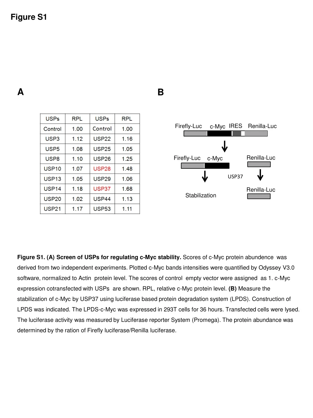
Figure S1 B A IRES Renilla-Luc Firefly-Luc c-Myc Renilla-Luc Renilla-Luc Firefly-Luc c-Myc USP37 Stabilization Figure S1. (A) Screen of USPs for regulating c-Myc stability. Scores of c-Myc protein abundence was derived from two independent experiments. Plotted c-Myc bands intensities were quantified by Odyssey V3.0 software, normalized to Actin protein level. The scores of control empty vector were assigned as 1. c-Myc expression cotransfected with USPs are shown. RPL, relative c-Myc protein level. (B) Measure the stabilization of c-Myc by USP37 using luciferase based protein degradation system (LPDS). Construction of LPDS was indicated. The LPDS-c-Myc was expressed in 293T cells for 36 hours. Transfected cells were lysed. The luciferase activity was measured by Luciferase reporter System (Promega). The protein abundance was determined by the ration of Firefly luciferase/Renilla luciferase.
Figure S2 B A Flag-USP37 - + - + - + - + HA-c-Myc + + + + Flag-Fbw7α - + - + Flag-USP37 - - + + siNC siFbw7 C siUSP37 - + - + HA-c-Myc Myc Flag-Fbw7α Flag-USP37 Actin Myc WT T58A S62A T58/S62A EGFP USP37 Myc/AA mutant Flag-USP37 EGFP Figure S2.The regulation of c-Myc by USP37 is independent of Fbw7. (A) The degradation of c-Myc by Fbw7 is blocked by USP37. 293T cells were transfected and protein levels were monitored by immunoblotting. (B) USP37 were co-expressed with wildtype c-Myc, c-MycT58A, c-MycS62A or c-MycT58/S62A in 293T cells and related proteins were detected using Western blotting. (C) Cells were transfected with siRNA against USP37 or Fbw7 as indicated. The protein levels were analyzed using Western blotting.
Figure S3 HA-MYC Flag-USP37 MERGE B A Figure S3. Colocalization of USP37 and c-Myc in the nucleus. (A) HA-c-Myc and Flag-USP37 were co-expressed into HeLa cells. Transfected cells were treated MG132, fixed, and the USP37 was stained with an anti-Flag antibody while c-Myc with anti-HA antibody. Images were analyzed by confocal microscopy. (B) HA-MAX or Flag-c-Myc were coexpressed with GFP-USP37 in HEK293T cells. Transfected cells were treated with MG132, and the cell lysates were immunoprecipitated using an anti-HA antibody.Co-immunoprecipitated USP37 was detected using an anti GFP-antibody. GFP-USP37 + + + + HA-MAX - + - + Flag-c-Myc - - + + IB:GFP IB:HA IP:HA IB:Flag IB:HA IB:Flag WCE IB:GFP
Figure S4 A B NS NS *** NS * Figure S4. Expression of USP37 in cancers. (A) The expression of USP37 is correlated with CCNB1. Scatter plots show a positive and significant correlation of mRNA expression between USP37 and the c-Myc target gene CCNB1 in lung adenocarcinoma. (B) mRNA expression data sets showing increased USP37 mRNA levels in breast invasive carcinoma(BRCA),but not in prostate adenocarcinoma(PRAD),head and neck squamous cell carcinoma(HNSC), kidney renal clear cell carcinoma(KIRC) or colon adenocarcinoma (COAD) compared to normal tissue.
Figure S5 USP37 A B Antibody absorbed Control USP37 DAPI MERGE Figure S5. Specificity of USP37 antibody. (A) H1299 cells were transfected with control or USP37 siRNA. USP37 was examined using immunofluorescence assay. (B) 0.4 mg USP37 antibody was incubated with 40 mg purified His-USP37 protein for 4 hours. Serial sections of lung cancer samples were stained with control antibody and the antibody absorbed by USP37 protein. siNC siUSP37