Download

1 / 14
140 likes | 161 Views
Segment medical images accurately, model mixtures of tissues, utilize Bayes theory, and enhance visualization through multi-dimensional Gaussian distributions and optimization techniques. This method enables more precise segmentation for various medical image sequences.

E N D
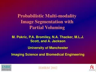
ISMRM 2002 Probabilistic Multi-modality Image Segmentation with Partial Voluming M. Pokric, P.A. Bromiley, N.A. Thacker, M.L.J. Scott, and A. Jackson University of Manchester Imaging Science and Biomedical Engineering
Problem Definition Segment the medical images to derive accurate and meaningful representation of all tissues present. The model is to be used in simulation and for visualisation (e.g. pre-operative planning, surgical rehearsal and training). ISMRM 2002 • Multi-dimensional image segmentation which models effect of mixtures of tissues present in a single voxel (i.e. partial volume effect). • Bayes theory used to obtain tissue probability maps to estimate the most likely tissue volume fraction present within each voxel.
Data Modelling Pure tissues - Gaussian distribution (blue lines). Mixtures of tissues - Triangular distribution convolved with Gaussian (red lines). The resulting distribution (green line).
Data Modelling Mt - mean tissue vector Ct - inverse of covariance matrix At - a constant which gives unit normalisation ISMRM 2002 A Multi-dimensional Gaussian Distribution for data gfor each tissue t
Bts- constant which gives unit normalisation h - fractional distance g along the line between two centres of distribution, h[0,1] N(g) – normal distance of g from the line between the two centres of distribution Tts(h) -partial volume distribution Ch - inverse covariance matrix: Ch= h Ct + (1-h) C A Multi-dimensional Partial Volume DistributionModelled along the line between two pure tissues means, Mtand Ms ISMRM 2002
Data Optimisation by Expectation Maximisation Expectation step - calculation of conditional probability of the model given the data using pure tissue and mixture of tissues distributions. ISMRM 2002
Data Optimisation by Expectation Maximisation Maximisation step - update of model parameters.
MR Image Sequences • IRTSE • 6850/18/300 • (TR/TE/TI) • TSE9 VE (PD) 5500/20 (TR/TE) TSE8 VE (T2) 5500/100 (TR/TE) TSE8 FLAIR 6000/100/2200 (TR/TE/TI) TSE19
Scatter Plots • Scatter plots for IRTSE and VE(PD) images for: • (a) original data • (b) density models with initial parameters • (c) density models after 10 iterations of EM algorithm (a) (b) (c)
Histogram Plots • Histogram plots for original data (red), sum of pure tissue models (green), sum of partial volume models (pink); sum of all models (blue) • (i)initial parameters (ii) parameters after 10 iterations
Histogram Plots • Histogram plots for original data (red), sum of pure tissue models (green), sum of partial volume models (pink); sum of all models (blue) • (i)initial parameters (ii) parameters after 10 iterations
Probability Maps Bone and air Soft tissue Fat WM CSF CSF GM
Half of the data we observed is due to partial voluming (mainly due to slice thickness, 3.5mm) Multi-dimensional segmentation with partial voluming enables more accurate segmentation of medical images of different modalities Better visual appearance of segmented tissues - important factors for simulation and visualisation This method can be applied to any sequence of images for which the linearity assumption holds ISMRM 2002 Conclusions
Acknowledgments AnIntegrated Environment for the Rehearsal and Planning of Surgical Interventions IERAPSI European Commission Project IST-1999-12175 http://www.tina-vision.net