Download
1 / 29
300 likes | 432 Views
Lower Leg, Ankle, and Foot Conditions. Chapter 16. Anatomical Review. Lower leg provides Support for the entire body Propulsion through space Adaptation to uneven terrain Absorption of shock. Forefoot. 5 metatarsals 14 phalanges

E N D
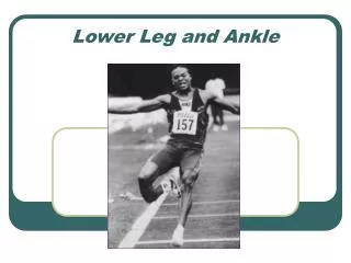
Lower Leg, Ankle, and Foot Conditions Chapter 16
Anatomical Review • Lower leg provides • Support for the entire body • Propulsion through space • Adaptation to uneven terrain • Absorption of shock
Forefoot • 5 metatarsals • 14 phalanges • Work together to form arches which distribute body weight throughout the foot • MTP-Metatarsophalangeal joints • IP-Interphalangeal • Phalanges work to transition weight from one foot to the other • Tarsometatarsal and Intermetatarsal joints • Allow foot to adapt to uneven surfaces during gait
Midfoot • Navicular • Bridges movements between the hindfoot and forefoot • Cuboid • 1st, 2nd, 3rd cuneiforms • Transverse tarsal joint • Calcaneocuboid (CC) lateral side • Talonavicular (TC) medial side • Permit only limited gliding
Midfoot continued… • Talocalcaneonavicular (TCN) joint • Allow gliding and rotation by a modified ball and socket joint • Plantar calcaneonavicular (spring) ligament inferiorly • Deltoid ligament medially • Bifurcate ligament laterally • Remaining midtarsal joints • Cuneonavicular, cuboideonavicular, cuneocuboid, and the intercuneiform
Hindfoot • Calcaneus • Anteromedial surface is the sustentaculum tali • Talus • Talus provides main articulation between the foot and the ankle • Articulations of the talus are • Talocrural • Subtalar
Talocrural • Modifies synovial hinge joint formed by the tibia, lateral malleolus of the fibula, and the talus • Fibula accounts for only 17% of the weighbearing • Lateral malleolus extends farther than medial which allows more inversion • Medial collateral ligament (deltoid) • Aterior tibiotalar (ATT) • Tibionavicular (TN) • Tibiocalcaneal (TC) • Posterior tibiotalar (PTT)
Talocrural continued… • Lateral ankle • Anterior talofibular (ATF) • Calcaneofibular (CF) • Posterior talofibular (PTF)
Subtalar Joint • Articulation between the facets of the talus and the sustentaculum tali on the superior calcaneus • Supported by • Intra-articular ligament • Talocalcaneal • Four small talocalcaneal ligaments
Tibioibular Joints • Proximal or superior joint is in the knee • Distal or inferior joint is supported by • Anterior and posterior tibiofibular ligaments • Allows some rotation • Some abduction or spreading • Space in-between is called the mortise
Plantar Arches • Longitudinal • Transverse • Primary supporting ligaments are: • Calcaneonavicular ligament (spring) • Long plantar ligament • Plantar fascia (plantar aponeurosis) • Short plantar ligament (plantar calcaneocuboid)
Muscles • Anterior compartment • Deep and superficial posterior compartment • Lateral compartment
Anterior compartment • Tibialis anterior • Extensor digitorum • Extensor hallucis longus • Peroneous tertius
Deep posterior compartment • Tibialis posterior • Flexor digitorum longus • Flexor hallucis longus
Superficial posterior compartment • Gastrocnemius • Soleus • Plantaris
Lateral compartment • Peroneus longus • Peroneus brevis
Muscles of the foot • Intrinsic- muscle has both attachments within the foot • Extrinsis- muscle has one attachment outside of the foot
Nerves • Sciatic • Tibial nerve (L4-S3) • Peroneal nerve (L4-S1) • Superficial peroneal nerve • Sural nerve
Kinematics • Toe flexion and extension • Flexor digitorum and hallucis • Extensor digitorum and halluci • Dorsiflexion • Tibialis anterior, extensor digitorum longus, and peroneous tertius • Plantarflexion • Soleus, gastrocnemius, plantaris, and flexor hallucis longus
Kinematics continued… • Supination • Calcaneal inversion, foot adduction and plantar flexion of the subtalar joint • Pronation • Calcaneal eversion, foot abduction, foot dorsiflexion
Toe and Foot Conditions • Hallus Rigidus • Hallus Valgus • Claw, hammer, and mallet toe • Turf toe • Reverse turf toe • Ingrown toenail • Metatarsalgia • Bunions • Retrocalcaneal bursitis
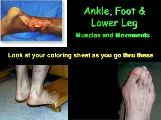
Contusions • Foot contusions • Lower leg contusions • Anterior compartment syndrome
Foot and Ankle sprains • Toe and Foot Sprains and dislocations • Lateral ankle sprains • Medial ankle sprains • Syndesmosis sprain • Subtalar sprain • Subtalar dislocation
Lower leg strains • Strains and tendinitis • Foot strains • Peroneal tendon strains • Tibialis posterior strain and rupture • Gastrocnemius strain • Achilles tendinitis • Achilles tendon rupture
Overuse Conditions • Plantar Fasciitis • Medial Tibial Stress Syndrome (MTSS) • Exertional Compartment Syndrome
Vascular and Neural Disorders • Venous disorders • DVT • Embolism • Plantar Interdigitial Neuroma • Tarsal Tunnel syndrome • Sural Nerve entrapment
Fractures • Freiberg’s disease • Sever’s disease • Stress fractures • Avulsion fractures • Osteochondral Fracture of the Talus
Displaced Fractures and Fracture Dislocations • Forefoot fractures • Tarsal fractures • LisFranc injury • Tibia-fibula fractures • Maisonneuve fracture • Ankle fracture-dislocations