Download
1 / 30
350 likes | 1.62k Views
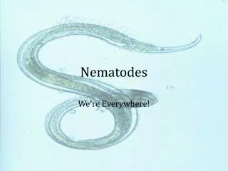
Nematodes . II BPT Dr Ekta Chourasia Lecturer, Microbiology . Nematodes – General Characters. Non-segmented cylindrical worms tapering at both ends Sexes are separate (diecious), male is smaller than female & its posterior end is curved ventrally Females are either

E N D
Nematodes II BPT Dr Ekta Chourasia Lecturer, Microbiology
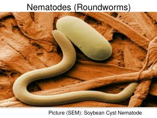
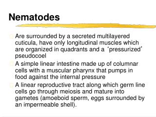
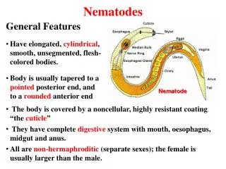
Nematodes – General Characters • Non-segmented cylindrical worms tapering at both ends • Sexes are separate (diecious), male is smaller than female & its posterior end is curved ventrally • Females are either • Viviparous (produce larvae/ embryos) • Oviparous (lay eggs) or • Ovo-viviparous (lay eggs which hatch immediately) • Live in intestinal tract or tissues Dr Ekta, Microbiology
Classification – Intestinal Nematodes Ascaris lumbricoides (round worm) Small Intestine only Necator americanus (american hook worm) Ancylostoma duodenale (hook worm) Strongyloides stercoralis Trichinella spiralis (trichina worm) Caecum and Vermiform appendix Enterobius vermicularis (pin worm) Trichuris trichiura (whip worm) Dr Ekta, Microbiology
Classification – Tissue Nematodes Lymphatic Wuchereria bancrofti Brugia malayi Brugia timori Subcutaneous Loa loa (african eye worm) Onchocerca volvulus (blinding filaria) Dracunculus medinensis (thread worm) Conjunctiva Loa loa Dr Ekta, Microbiology
Modes of Infection of Nematodes • Ingestionof – • Embryonated eggs contaminating food & drinks, e.g. A.lumbricoides, E. vermicularis & T. trichiura • Growing embryos in an intermediate host (infected cyclops) e.g. D.medinensis • Encysted embryos in infected pig’s flesh e.g. Trichinella spiralis • Penetration of skin – filariform larvae bores through the skin e.g. A.duodenale, S.stercoralis, N.americanus • By blood sucking insects e.g. filarial worms • Inhalation of infected dust containing embryonated eggs e.g. A.lumbricoides, E.vermicularis Dr Ekta, Microbiology
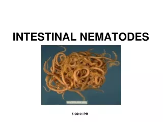
INTESTINAL NEMATODES Dr Ekta, Microbiology
Ascaris lumbricoides (roundworm) Adult worms Male 15 to 30 cms Female 20 to 40 cms, oviparous Eggs 60 µ, bile stained Albuminous coat with unsegmented ovum Infective form Embryonated eggs Mode of transmission Ingestion Site of localization Small intestine Dr Ekta, Microbiology
Pathogenicity & Clinical Features • Ascariasis – infection of A.lumbricoides • Symptoms divided into two groups: those produced by • Migrating larvae – pneumonia (loeffler’s syndrome), Visceral larva migrans • Adult worms Dr Ekta, Microbiology
Symptoms & Complications • Symptoms produced by Adult worms • Abdominal discomfort, anorexia, nausea & diarrhoea. • PEM, Vit. A deficiency (night blindness) • Intestinal obstruction (particularly in children 1-5 years), intussusception & volvulus • Penetration through intestinal ulcer (perforation) – peritonitis • Hypersensitivity reactions to worm Ags (toxic body fluids) – urticaria, edema of face, conjunctivitis, irritation of URT • Ectopic Ascariasis Dr Ekta, Microbiology
Laboratory Diagnosis • Macroscopic - Direct detection of worm/s in stool or vomit • Microscopic – direct examination of feces following floatation method: bile stained eggs.(eggs may not be seen at least 40 days after infection) • Blood examination – eosinophilia. Dr Ekta, Microbiology
Treatment Mebendazole/ Albendazole – drug of choice but contraindicated in pregnancy & heavy infection Pyrantel pamoate – single dose Piperazine citrate Levamisole Prevention Good sanitation and personal hygiene Mass treatments with single dose mebendazole or albendazole for all school-age children every three to four months Treatment & Prevention Dr Ekta, Microbiology
Ancylostoma duodenale (hook worm) Adult worms Male 8 -11mm Female 10-13 mm, oviparous Eggs 60 µ, non bile stained (colorless) Segmented, 4 blastomeres Infective form 3rd stage filariform larva Mode of infection Penetration into skin Site of localization Small intestine Dr Ekta, Microbiology
6 to 8 weeks 8 to 10 days 48 hours 6 to 8 days Life cycle of hookworm Dr Ekta, Microbiology
Hook worms in the intestine Dr Ekta, Microbiology
Pathogenicity & Clinical Features • Ancylostomiasis or hookworm disease, characterised by iron deficiency anaemia • Ancylostome dermatitis or Ground itch • Creeping eruption • Bronchitis & bronchopneumonia. • Abnormal appetite showing Pica or Geophagy – perverted taste for earth, mud or lime • Growth retardation • General appearance – pale plumpy with protuberant abdomen & dry lustreless hair. Dr Ekta, Microbiology
Laboratory Diagnosis • Stool examination – microscopy: non bile stained egg, segmented • Occult blood in stool – positive • Blood examination – anaemia, eosinophilia Dr Ekta, Microbiology
Strongyloides stercoralis Adult worms 2 - 2.5mm, ovoviviparous, eggs laid in the tissues Free living worms Moist soil Infective form Filariform larvae Mode of transmission Penetration / autoinfection Site of localization Wall of Small intestine, mainly duodenum & jejunum Dr Ekta, Microbiology
Pathogenicity • Diseases • Dermatitis, erythema, itching • Creeping eruption / larva migrans • Bronchopneumonia / malabsorption • Brain abscess, meningitis, peritonitis - compromised (Hyper infection) • Diagnosis - rhabditiform larva in stool • Treatment - Thiabendazole / Mebendazole Dr Ekta, Microbiology
Trichinella spiralis (Trichina Worm) Adult worms (smallest nematode infecting man) Male 1.4 – 1.6 mm Female 3 - 4 mm, viviparous Encysted larvae (100µ) in striated muscles of pig Infective form Mode of transmission Ingestion of improperly cooked pork Site of localization Small intestine Diaphragm, Intercostals, Deltoid, Pectoralis major, Biceps Commonly involved muscles Dr Ekta, Microbiology
Pathogenicity & Diagnosis • Disease - Trichinelliasis / Trichinosis • GI disturbances • Muscle swelling & weakness • Complications – myocarditis, encephalitis • Lab diagnosis • Muscle biopsy – encysted larva • Blood – eosinophilia between 2nd & 4th week • Serology – to detect specific Abs Dr Ekta, Microbiology
Treatment Thiabendazole & Mebendazole – adult worms Corticosteroids – complications Prevention Proper cooking of pork or proper storage Avoidance of feeding bits & refuse from slaughter houses & farms to pigs – breaks life cycle. Treatment & Prevention Dr Ekta, Microbiology
Enterobius vermicularis (Pin Worm, Seatworm) Adult worms Male 2 - 5 mm Female 8 -13 mm, oviparous 60 µ, non bile stained Plano-convex with coiled embryo Eggs Infective form Embryonated egg Mode of transmission Ingestion, Autoinfection Site of localization Large intestine – caecum & appendix Dr Ekta, Microbiology
Life cycle – E. vermicularis Dr Ekta, Microbiology
Clinical features • Due to migration of worm - Perianal, perineal & vaginal itching (pruritis) worsens at night. • Insomnia and restlessness • Nocturnal enuresis Dr Ekta, Microbiology
Laboratory Diagnosis & Treatment • Detection of adult worms in- • Feces • Perianal region • NIH swab – scrapings from perianal region • Microscopy – non bile stained eggs • Mebendazole, pyrantel pamoate Dr Ekta, Microbiology
Trichuris trichiura (Whip Worm) Adult worm 30 – 50 mm Eggs 60 µ, bile stained Barrel-shaped with Mucus plug at each pole Unsegmented ovum Infective form Mature embryonated eggs Mode of transmission Ingestion Site of localization Large intestine - caecum Dr Ekta, Microbiology
Clinical features • Infection – Trichuriasis • chronic profuse mucus and bloody diarrhea with abdominal pains and edematous rectum • malnutrition, weight loss and anemia Dr Ekta, Microbiology
Laboratory diagnosis & Treatment • Stool examination – bile stained eggs with bipolar mucus plugs • Treatment – albendazole / mebendazole • Prevention – • Proper disposal of night soil • Prevention of consumption of uncooked vegetables & fruits . Dr Ekta, Microbiology
Key to the diagnosis of Intestinal Nematodes Dr Ekta, Microbiology