Download
1 / 26
430 likes | 1.71k Views
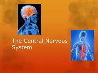
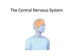
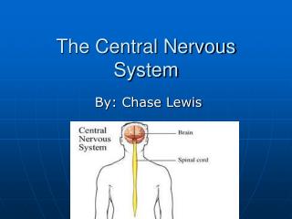
The Central Nervous System. The CNS consists of the brain & the spinal cord , immersed in the cerebrospinal fluid (CSF) The brain consists of three main structures: the cerebrum , the cerebellum & the brainstem

E N D
The Central Nervous System • The CNS consists of the brain & the spinal cord, immersed in the cerebrospinal fluid (CSF) • The brain consists of three main structures: the cerebrum, the cerebellum & the brainstem • Cerebrumcomposed of 2cerebral hemispheres each made up of the cerebral cortex (grey matter), the basal nuclei & diencephalon (white matter) • Each hemisphere (left and right), consists of four lobes (frontal, parietal, occipital & temporal)
cerebral cortex • The outer layer of grey matter, approximately 2 mm thick, covering the entire surface of the cerebral hemispheres • It is made up of neurons & supporting glial cells • It functions to correlate information from many sources to control cognitive function (all aspects of perceiving, thinking & remembering)
The basal nuclei is a group of cell bodies deep in the white matter of the cerebral hemispheres beneath the cerebral cortex Major components include the caudate, putamen, globus pallidus &substantia nigra The basal nuclei functions to sensorio-motor integration & motor control Basal Nnuclei (Ganglia)
Cerebellum • The walnut-shaped structure situated at the base of the brain • The cerebellum is responsible for motor co-ordination, posture & maintaining equilibrium • It ordinates sensory input from the inner ear and the muscles to provide accurate control of position & movement
Brain Stem • The stalk-like part of the brain connecting the cerebral cortex, white matter & the spinal cord • Made up of the pons, the medulla oblongata & the midbrain • The brainstem acts as an important relay station • The brainstem contributes to the control of breathing, sleep & circulation • Every nerve impulse passing between the brain & the spinal cord must pass through the brainstem to allow the body to function normally
Thalamus and Hypothalamus The thalamus has wide connections with the cortex the basal ganglia, hypothalmus & brainstem It is capable of perceiving pain The hypothalamus has several functions, including control of the body’s appetite, sleep patterns, sexual drive & response to anxiety Ventricles Ventricles are a number of cavities within the brain Ventricles are filled with CSF, which is produced within the ventricle wall The CSF also surrounds the outer surfaces of the brain and ‘cushions’ the brain against trauma, maintains and control the extracellular environment, and circulates endocrine hormones Other Parts of the Brain
The limbic system • It a group of nerve pathways including structures deep within the temporal lobes, as thehippocampus& the amygdale • It is connected with the cerebral cortex, white matter & brainstem • the limbic system is involved in the control and expression of mood and emotion, in the processing and storage of recent memory, and in the control of appetite and emotional responses to food • The limbic system has been implicated in the pathogenesis of depression & schizophrenia • The limbic system is linked with parts of the neuro-endocrine & autonomic nervous systems hippocampus
Reticular Activating System • It is a collection of nuclei at the core of the brainstem • They receive input from the body’s sensory systems (sight, smell, taste, …etc), the cerebellum and cerebral hemispheres • Some neurons from the reticular formation project to meet motor neurons of the spinal cord and influence functions like CVS & respiratory control • In addition, there are also neurons projecting into most of the rest of the brain • The ascending fibers of the reticular formation form a network called the reticular activating system, which influence wakefulness, overall degree of arousal and consciousness (depression!)
Neurotransmission & Processing of Information • Two signaling mechanisms; action potentials & synaptic signals are the basis for all the information-processing in the brain • An action potential is initiated at a synapse and travels along the axon to the axonal terminal • The electrical signal is converted to a chemical signal; a neurotransmitter • It diffuses out of the neuron, across the synapse, to its neighboring neuron • At the postsynaptic neuron the chemical signal is converted back into an electrical signal once again
The Action Potential • Resting neurons have a negative membrane potential, caused by a steady outflow of potassium ions & an impermeability to sodium ions • The action potentialrepresents transient changes in this resting membrane potential • For most axonsde-polarisation initiates the action potential • It causes a transient change in the membrane allowing the passage of sodium ion
The Synaptic Signal 50nm • Once the action potential reaches the axonal terminal, the changed membrane potential triggers the activation of calcium channels • This allows elevation of the concentration of calcium ions in the pre-synaptic neuron • Thereafter, synaptic vesicles fuse with the presynaptic membrane and one or more neurotransmitters are released into the synaptic cleft • Neurotransmitters bind to specific receptors on the membrane of the postsynaptic neuron, or to an autoreceptor
Neuropeptide Neurotransmitters • Corticotropin releasing hormone • Corticotropin (ACTH) • Beta-endorphin • SubstanceP • Neurotensin • Somatostatin • Bradykinin • Vasopressin • Angiotensin II
Serotonin • CNS contains only less than 2% of the total serotonin in the body • Serotonin is localized mainly in nerve pathways emerging from the raphe nuclei, a group of nuclei at the centre of the reticular formation • These serotonergic pathways spread extensively throughout the brainstem, the cerebral cortex and the spinal cord • In addition to mood control, serotonin has other functions, including the regulation of sleep, pain perception, body temperature, blood pressure & hormonal activity
Serotonin Receptors • Seven classes, 5-HT1- 5-HT7 • All 5-HT1A,1B,1D,1E,1F are inhibitory Gi-coupled • 5-HT1Ais a somatodentritic auto-receptor • 5-HT1B,1D are postsynaptic & pre-synaptic auto-Rs • 5-HT2 & 5-HT4 are excitatory receptors linked to Gq/11-PLC and Gs-AC respectively • 5-HT3 is the only inotropic 5-HT receptor
Noradrenaline • Noradrenergic neurons are found in the locus coeruleus, the pons & the reticular formation • These neurons project to the cortex, hippocampus, thalamus & midbrain • The release of NA tends to increase the level of excitatory activity within the brain • Noradrenergic pathways are thought to be involved in the control of functions such as attention & arousal locus coeruleus: A small area in the brainstem consisting of a pair of identical nuclei in the pons from which all brain connections, using NA, arise
Dopamine It is of high density in the basal ganglia Dopaminergic neurons are widely distributed throughout the brain in three important dopamine pathways: The nigrostriatal, The mesocorticolimbic, The tubero-hypophyseal DA deficit in Parkinson’s disease, DA over activity inschizophrenia Acetylcholine Cholinergic pathways are concentrated mainly in the brainstem They are believed to be involved incognitivefunctions, especially memory Severe damage to these pathways is the probable cause of Alzheimer’s disease Dopamine & Acetylcholine
Dopamine Receptors • DA receptors are 7-TM G-protein coupled • D1-like(D1, D5) receptors are linked to Gi-AC • D1-like receptors mediate excitatory DA activity • D5 is abundant in limbic system, D1 in striaum, little in limbic system • D2-like(D2, D3, D4) are linked to Gs-AC • D2-like receptors mediate inhibitory activity of DA • D3, D4 receptors are located mainly in limbic s, & not in motor structures, unlike D2 receptors • Atypical neuroleptics (clozapine) have affinity of D3, D4> D2 → less extra-pyramidal side effects
Exciatatory Amino Acid NeurotransmissionGlutamate & Aspartate • Exciatatory amino acid neurotransmitters, especially glutamate, are abundant in the CNS • Glutaminergic neurons in cerebral cortex provide the main excitatory cortical output to hippocampus, basal ganglia, thalamus & amygdala • Hippocampal glutaminergic neuronsproject to limbic structures possibly controling learning • Retinal glutaminergic neuronsare the major excitatory pathways linked to photoreceptors • NMDA receptor antagonists including glycine-binding site blockers are investigated as potential antiepileptic agents or drugs that prevent ischemic brain damage after stroke/trauma
NMDA Receptors Inotropic tetrameric receptor permeable to monovalent cations, high permeability to Ca2+ Binding to glutamate→ ↑Ca2+ → Activation of Ca2+-dependent enzymes (PKC, NOS)→ response It has 6 binding sites, 2 excitatory: Glutamate binding sites Glycine binding sites and 4 inhibitory sites Phencyclidine (PCP), voltage-gated Mg2+, Zinc, and polyamine binding site Non-NMDA Receptors 4 subtypes named after their selective agonist: Kainate R; Na+, K+-inotropic receptor, abundant in hippocampus Quisqualate/AMPA iontropic Na+, K+- regulating receptor L-AP4 metabotropic mGluR4,6,7,8 presynaptic receptors They cause inhibition of pre-synaptic neurons ACPD metabotropic receptors linked to PLC mediating excitatory effects Exciatatory Amino Acid ReceptorsNMDA (N-Me-D-aspartate) & Non-NMDA Receptors
GABA, the main inhibitory transmitter CNS in GABA pathways & GABA inter-neurones 50% of the inhibitory synapses in the brain are GABA mediated Synthesized by glutamate decarboxylation Pre- & post-synaptic GABA transportersterminate effect GABAergic inter-neurons in retina, cortex, hippocampus, spinal cord, & cerebellum Inhibitory Amion Acid TransmissionGama-aminobutyric Acid (GABA)
Inhibitory Anion Acid TransmissionGlycine • Formed from serine by hydroxyMe-transferase • Glycine receptors are inotropic Cl--channels, blocked by strychnine • Glycine is removed by uptake using two transporters GLYCT-1 & -2 • Limited distribution • in spinal cord as inhibitory control over motor neurons • In brainstem, reticular formation & retina
Neuropeptides • Most important ones include: opioid peptides, angiotensin II, oxytocin, cholecystokinin, and vasopressin • All aforementioned peptides are excitatory except for opioid peptides that are inhibitory • Opiods will be discussed in detail later in separate session