Download
1 / 115
1.16k likes | 1.17k Views
Fluid Balance & Acute Kidney Injury Study Package . This study package has been designed to aid multidisciplinary staff in developing their knowledge of Acute Kidney Injury and Fluid Balance.

E N D
This study package has been designed to aid multidisciplinary staff in developing their knowledge of Acute Kidney Injury and Fluid Balance. The best format for this package is an interactive study day with a facilitator and expert faculty members to deliver the content. Any reference to patients and blood results are fictional and confidentiality is maintained at all times.
Objectives: • To develop a basic understanding of what the kidneys do and how they work at cellular level. • Gain further knowledge in understanding the biochemistry that relates to AKI. • Discuss the common causes of AKI and how these may be treated. • Understand some of the common medications associated with AKI. • Discuss the concept of fluid balance and the challenges we face to get this right. • Consolidate learning through case scenario discussions.
References • The Mid Staffordshire NHS Foundation Trust Public Inquiry (2013) Report of the Mid Staffordshire NHS Foundation Trust Public Inquiry: executive summary. London:StationeryOffice (Chair: R Francis). Available at: www.midstaffspublicinquiry.com/sites/default/files/report/Executive%20summary.pdf • A review of the care of patients who died in hospital with a primary diagnosis of acute kidney injury (acute renal failure). National Confidential Enquiry into Patient Outcome and Death (NCEPOD). 2009
Objectives: • To develop a basic understanding of what the kidneys do and how they work at cellular level. • Gain further knowledge in understanding the biochemistry that relates to AKI. • Discuss the common causes of AKI and how these may be treated. • Understand some of the common medications associated with AKI. • Discuss the concept of fluid balance and the challenges we face to get this right. • Consolidate learning through case scenario discussions.
Functions of the Kidney • Regulation of electrolytes • Maintenance of acid–base balance • Regulation of blood pressure (via maintaining salt and water balance). • Natural filter of the blood, and removes water soluble wastes, which are diverted to the urinary bladder. • Production of Urine • Reabsorption of water, glucose, and amino acids. • The kidneys also produce hormones including calcitriol, erythropoietin, as well as the enzyme renin.
Components of the Body regulated by the kidneys Electrolytes: Na, K, Chloride Total Body Water Ph: By excreting hydrogen ions By regulating the concentration of HCO3- the major extracellular buffer Minerals: Calcium, Phosphurus and Magnesium Endogenously produced waster products: Urea-end point of protein catabolism Creatinine-produced by skeletal muscle Uric Acid-nucleic acid breakdown product
Endocrine Functions • Sole source of Erythropoietin (red blood cell production) • Produces final enzyme to produce Vitamin D • Produces Renin • Paracrine Substances within the kidney such as prostaglandins and endothelin which are produced in response to injury and may act as vasoconstrictors
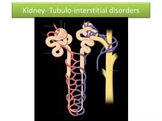
Nephron • Each kidney contains 1 million nephrons • Functional Unit of the Kidney • Blood is first filtered and then different components reabsorbed or further secreted along tubules • The resulting fluid is passed from the kidneys down the ureters to the bladder and is excreted as urine
Autoregulation • Blood flow directly affects the glomerulus filtration rate • The blood flow into the glomerulus is via the afferent arteriole and the blood flow out of the glomerulus is by the efferent arteriole • Altering the radius of both or one of these vessels will alter the pressure within the glomerulus.
The renin angiotensin aldosterone system is a series of reactions designed to help regulate blood pressure. • When blood pressure falls (for systolic to 100mmhg or lower) the kidneys release the enzyme renin into the bloodstream. • Renin splits angiotensinogen, a large protein that circulates in the bloodstream into pieces. One piece is angiotensin I. • Angiotensin I (relatively inactive) is split into pieces by angiotensin-converting enzyme (ACE). One piece is angiotensin II: a hormone which is very active. • Angiotensin II causes the muscular walls of efferent arterioles to constrict increasing blood pressure. It also triggers the release of aldosterone (a hormone) from the adrenal gland and antidiuretic hormone from the pituitary gland. • Aldosterone and antidiuretic hormone cause the kidneys to retain salt. Aldosterone can also cause the kidneys to excrete potassium. The increased sodium causes water to be retained thus increasing blood volume and blood pressure.
Water Haemostasis • Water balance is controlled by antidiuretic Hormone (ADH). • ADH is released in response to three stimuli: -Increased blood osmolality (concentration of blood constituents) -Decreased blood volume -Angiotension II • Receptors facilitate greater water reabsorbtion.
Acid Base Balance • Maintenance of a constant pH is important as many of our enzymes are pH sensitive. • pH is the concentration of hydrogen ions • Bicarbonate is the main buffer to acid in the human body and is filtered by the kidneys so must be reabsorbed. • H2O + CO2 HCO3- + H+
Summary • The kidneys play a vital role in maintaining blood volume. • Blood flow into the kidneys maintains function. • Any alterations in kidney pathology could result in acid base imbalance, electrolyte disturbance and interruption to regulation of blood pressure.
References • renin-angiotensin-aldosterone system http:// www.merckmanuals.com/home/heart_and_blood_vessel_disorders/high_blood_pressure/high_blood_pressure.html
Objectives • To develop a basic understanding of what the kidneys do and how they work at cellular level. • Gain further knowledge in understanding the biochemistry that relates to AKI. • Learn the common causes of AKI and how these may be treated. • Discuss the concept of fluid balance and the challenges we face to get this right. • Consolidate learning through case scenario discussions.
The kidney and biochemistry • Three major functions • Excretion of waste • Maintenance of fluid balance • Hormone synthesis
Sodium (134-145mmol/L) • Most abundant extracellular cation • Carries a positive charge • Important in determining water distribution across cell membranes • Input and output usually balanced • 25000mmol/L filtered at the glomerulus • Normally 99% reabsorbed
Sodium and water control • Sodium • Regulated by aldosterone • Adrenal glands • Low sodium, aldosterone production increases to increase renal reabsorption (in exchange for potassium/hydrogen) • Water • Regulated by Vasopressin (Anti-diuretic hormone) • Hypothalamus • Stimulated by rising osmolality, low circulating blood volume • Increased water reabsorption by renal collecting ducts
Potassium (3.5-5.3 mmol/L) • Main intracellular cation • 98% is stored within cells • Reasons for movement out of cell • Acidosis • Lack of insulin • Severe cell damage • External balance determined by intake and renal excretion • Plasma K+ poor indicator of body content • However, it is the changes in the extracellular concentration that affect neuromuscular and cardiac function
Chloride (98-108 mmol/L) • Extracellular anion • Sodium and chloride involved in maintenance of water distribution • Filtered at the glomeruli and reabsorbed in proximal tubule • Changes should mirror sodium
Urea (2.5-7.8 mmol/L) • Synthesised in liver • By-product of the deamination of amino acids • Elimination in urine is major route for nitrogen excretion • Filtered from blood at the glomerulus • Significant tubular reabsorption
Creatinine (50-110 mmol/L) • Most reliable biochemical test of glomerular function • End product of nitrogen metabolism • Changes can occur independently of renal function • Muscle mass changes • Immediately after surgery • Steroid treatment • Re-feeding
GFR – Glomerular filtration rate • Reflects the number of functioning glomeruli • Estimate of renal impairment • Serum sample for U&E’s • Calculation of glomerular filtration rate using the following formula: • 186 x (Creat / 88.4)-1.154 x (Age)-0.203 x (0.742 if female) x (1.210 if black) eGFR should not be used in identifying Acute Kidney Injury but for monitoring chronic kidney disease and function.
GFR – Glomerular filtration rate • More reliable that creatinine clearance • Removes inaccuracies of urine collections • Separate formula for children and those with renal failure
Blood gases – acid base • Kidneys vital for the excretion of Hydrogen ions (H+) • If increase in H+ acidosis (Low pH) • If decrease in H+ alkalosis (High pH) • Kidneys not functioning means the patient is at risk of acid-base disorder.
Urinalysis • Provides important information about kidney function • Combur 7 • Seven patch test strip • Visually read or using urisys meter • Fresh samples only • Mix well before use
Tests • pH • Glucose • Ketones • Leucocytes • Nitrites • Protein • Blood
Interpretation • pH • acid base status of urine • alkaline pH indicates old sample or urinary tract infection • Protein • presence suggests renal disease • Glucose • Generally found in urine at blood concentrations >10 mmol/L • Can suggest diabetes mellitus • Reduced renal absorption
Interpretation • Blood • red blood cells, hemoglobin, or myoglobin (muscle hemoglobin) • sensitive early indicator of renal disease • Ketones • normal product of fat metabolism • increased amounts seen in diabetes or starvation (extreme dieting) • Nitrites • certain bacteria convert normal urine nitrate to nitrite • indicator of urinary tract infection • Leucocytes • indicator of urinary tract infection
Levels of detection • Just because there is an analyte detected does not mean that there is underlying pathology • Use locally derived action limits • Action limit for protein = 30mg/dL
Limitations • Captopril, phenoketones can produce false positive ketone results • Imipenen, meropenem and clavulanic acid can produce false positive leucocytes • False positive blood results 3 days before and 3 days after period
Good Practice • Analyse sample as soon as possible • Thoroughly mix the sample • Wear PPE • Note smell, colour and clarity • Analyse in well lit area • Dip the reagent strip into the specimen ensuring all areas are covered. Remove after 2 seconds • Tap the edge of the strip to remove excess urine • If test to be read visually wait 2 minutes before reading strip • Ensure results transferred to notes
Laboratory results and AKI • Since March 2015 all NHS trusts in England should have implemented a national algorithm in their biochemistry departments for Standardising the early detection of AKI. (NHS England 2015) • This laboratory system will generate an AKI result and score (1,2 or 3) depending on previous creatinine results. • It is essential that your clinical assessment is used in conjunction with the blood result to formulate the diagnosis. • Urine output is also a key indicator of acute kidney injury and must be considered. • Pregnancy, extremes in muscle mass, CKD patients may generate false positive results. CAREFUL CLINICAL ASSESSMENT IS VITAL.
Responsibility • If blood samples have been taken ensure results are reviewed! • Medics • Ensure you are aware of patients recent blood results and trends that may be developing • Pharmacists • When undertaking a medication review ensure you have access to patient results • Nursing team • If you are aware of abnormal results ensure they are being acted upon
Laboratory contact • Phone wards to inform users of critically abnormal results • Follow the FRCPath recommendations and locally agreed guidance • Sodium <120 >155 • Potassium <2.7 >6.5 • Urea >25 (if no previous abnormal urea within current admission) • Creatinine >400 (if no previous abnormal)
Summary • Rise in creatinine is the only laboratory test that can aid in the diagnosis of AKI. • Ensure blood results requested are reviewed • Ensure action is taken and documented • Any clinical questions contact Clinical Scientist • Any diagnosis of AKI should be made in conjunction with cautious assessment, patient history and clinical examination.