Download
1 / 45
450 likes | 464 Views
This article discusses the differences between the Somatic Nervous System (SNS) and Autonomic Nervous System (ANS), including their functions, anatomy, and organization. It also explores the divisions within the ANS and the role of the enteric nervous system.

E N D
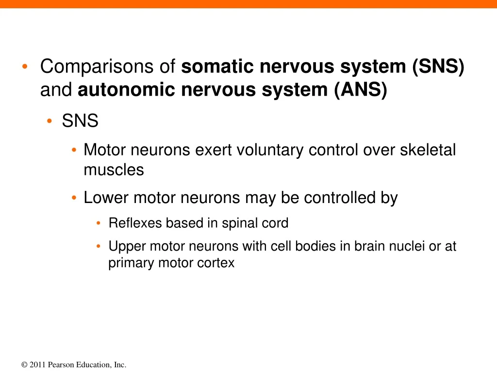
Comparisons of somatic nervous system (SNS) and autonomic nervous system (ANS) SNS Motor neurons exert voluntary control over skeletal muscles Lower motor neurons may be controlled by Reflexes based in spinal cord Upper motor neurons with cell bodies in brain nuclei or at primary motor cortex
A schematic of the somatic nervous system (SNS), which provides conscious and sub- Conscious control over skeletal muscles Upper motor neurons in primary motor cortex BRAIN Somatic motor nuclei of brain stem Skeletal muscle Spinal cord Lower motor neurons Somatic motor nuclei of spinal cord Skeletal muscle
Section 1: ANS Functional Anatomy and Organization Comparisons of somatic nervous system (SNS) and autonomic nervous system (ANS) (continued) ANS CNS motor neurons synapse with visceral motor neurons in autonomic ganglia, which control visceral effectors Integrative centers located in hypothalamus Two types of visceral motor neurons Preganglionic neurons (cell bodies in CNS) Activities represent direct reflex responses Ganglionic neurons (cell bodies in autonomic ganglia) Innervate effectors like cardiac and smooth muscle
Visceral motor nuclei in hypothalamus BRAIN Preganglionic neuron Visceral Effectors Smooth muscle Autonomic nuclei in brain stem Autonomic ganglia Glands Ganglionic neurons Spinal cord Cardiac muscle Autonomic nuclei in spinal cord Adipocytes Preganglionic neuron A schematic of the autonomic nervous system (ANS), which controls visceral functions largely outside our awareness
Module 14.1: ANS divisions Three ANS divisions Sympathetic (or thoracolumbar) division Axons emerge from thoracic and superior lumbar segments of spinal cord Innervate ganglia relatively close to spinal cord “Kicks in” only during periods of exertion, stress, or emergency Parasympathetic (or craniosacral) division Axons emerge from brain stem and sacral spinal segments Innervate ganglia very close (or within) target organs Most often, effects are opposite to sympathetic
Autonomic Nervous System Sympathetic Division Parasympathetic Division In the sympathetic division, or thoracolumbar (thor-a-kō-LUM-bar) division, axons emerge from the thoracic and superior lumbar segments of the spinal cord and innervate ganglia relatively close to the spinal cord. In the parasympathetic division, or cranio- sacral (krā-nē-ō-SĀ-krul) divions, axons emerge from the brain stem and the sacral segments of the spinal cord, and they innervate ganglia very close to (or within) target organs. Cranial nerves (III, VII, IX, and X) T1 T2 T3 T4 The two main divisions of the ANS: the sympathetic and parasympathetic divisions T5 T6 Thoracic nerves T7 T8 T9 T10 T11 T12 L1 Lumbar nerves (L1, L2 only) L2 S2 Sacral nerves (S2, S3, S4 only) S3 S4
Module 14.1: ANS divisions Three ANS divisions (continued) Enteric nervous system (ENS) Extensive network of neurons (~100 million) and nerve networks within walls of digestive tract Influenced by sympathetic and parasympathetic divisions However, many complex visceral activities are coordinated on a local level (without CNS instructions) Will be discussed more in Digestive System
The extensive system of neurons and nerve networks of the enteric nervous system (ENS) Esophagus Stomach Large intestine Small intestine
Aortic arch Right vagus nerve Trachea Autonomic Plexuses and Ganglia Left vagus nerve Cardiac plexus Thoracic spinal nerves Pulmonary plexus Esophagus Thoracic sympathetic chain ganglia Esophageal plexus Splanchnic nerves Celiac plexus and ganglion Superior mesenteric ganglion Diaphragm Superior mesenteric artery Inferior mesenteric plexus and ganglion Inferior mesenteric artery Hypogastric plexus Pelvic symapthetic chain Representative plexuses, ganglia, and nerves of the sympathetic and parasympathetic divisions
Autonomic ganglia Sympathetic division Preganglionic fibers (neurons) are relatively short while postganglionic fibers (neurons) are relatively long Accordingly, sympathetic ganglia (where these fibers synapse) are relatively near spinal cord Specific ganglia Sympathetic chain (on either side of spinal cord) Innervates visceral effectors in thoracic cavity, head, body wall, and limbs
Sympathetic division (continued) Specific ganglia (continued) 2.Collateral ganglia (within abdominopelvic cavity) Includes celiac, superior, and inferior mesenteric ganglia Innervates visceral effectors in abdominopelvic cavity Adrenal medulla Center of adrenal gland Acts as endocrine gland Targets organs and systems throughout body
Module 14.2: Autonomic ganglia Sympathetic division (continued) Prepares body for heightened levels of somatic activity Known as “fight or flight” division Typical responses Heightened mental alertness Increased metabolic rate Reduced digestive and urinary functions Activation of energy reserves Increased respiratory rate and dilation of passageways Elevated heart rate and blood pressure Activation of sweat glands
Organization of the sympathetic division of the ANS KEY Preganglionic fibers Postganglionic fibers Ganglionic neurons in the sympathetic chain and collateral ganglia exert their effects through innervation of peripheral target organs. Hormones released into circulation Target Organs Ganglionic Neurons Each sympathetic chain consists of a series of interconnected ganglia located on either side of the vertebral column. Visceral effectors in thoracic cavity, head, body wall, and limbs Preganglionic Neurons Lateral gray horns of spinal segments T1-L2 The collateral ganglia, located within the abdominopelvic cavity, include the celiac, superior mesenteric, and inferior mesenteric ganglia. Visceral effectors in abdomino- pelvic cavity Organs and systems throughout the body The center of each adrenal gland contains a sympathetic ganglion, the adrenal medulla, that acts as an endocrine organ. Ganglionic neurons in the adrenal medullae affect target organs throughout the body through the release of hormones into the general circulation.
Parasympathetic division Typical preganglionic fiber synapses on 6–8 ganglionic neurons May be situated in: Terminal ganglia (near target organ) Usually paired Ciliary ganglion (intrinsic eye muscles) Pterygopalatine and submandibular ganglia (nasal, tear, and salivary glands) Otic ganglion (parotid salivary gland)
Parasympathetic division (continued) Typical preganglionic fiber synapses on 6–8 ganglionic neurons (continued) May be situated in: (continued) Intramural (murus, wall) ganglia (embedded in target organ wall) Typically interconnected masses/clusters of cells Innervate visceral organs of neck, and of thoracic and abdominopelvic cavities
Module 14.2: Autonomic ganglia Parasympathetic division (continued) Concerned with regulation of visceral function and energy conservation Known as “rest and digest” system Typical responses Decreased metabolic rate Decreased heart rate and blood pressure Increased salivary and digestive gland secretion Increased digestive tract motility and blood flow Stimulation of urination and defecation
Organization of the parasympathetic division of the ANS KEY Preganglionic fibers Postganglionic fibers Preganglionic Neurons Ganglionic Neurons Target Organs III Intrinsic eye muscles (pupil and lens shape) Ciliary ganglion The midbrain, pons, and medulla oblongata contain parasympathetic nuclei associated with cranial nerves III, VII, IX, and X. VII Pterygopalatine and submandibular ganglia Nasal glands, tear glands, and salivary glands IX X Otic ganglion Parotid salivary gland Visceral organs of neck, thoracic cavity, and most of abdominal cavity Intramural ganglia In sacral segments of the spinal cord, parasympathetic nuclei lie in the lateral gray horns of spinal segments S2–S4. Visceral organs in inferior portion of abdominopelvic cavity Pelvic nerves Intramural ganglia
Module 14.3: Autonomic innervation patterns Autonomic innervation patterns Sympathetic division Every spinal nerve has a gray ramus carrying sympathetic postganglionic fibers Preganglionic fibers passing to collateral ganglia form splanchnic nerves Postganglionic fibers innervating thoracic cavity structures form bundles or sympathetic nerves
Module 14.3: Autonomic innervation patterns Parasympathetic division Vagus nerve (X) alone provides ~75% of all parasympathetic outflow Numerous vagus nerve branches intermingle with sympathetic fibers forming nerve plexuses Preganglionic fibers in sacral spinal cord segments form distinct pelvic nerves Innervate intramural ganglia in kidneys, bladder, terminal portions of large intestine, and sex organs
The innervation of the parasympathetic division on one side of the body; the innervation on the opposite side (not shown) follows the same pattern KEY Preganglionic neurons Ganglionic neurons Pterygopalatine ganglion Lacrimal gland Eye Ciliary ganglion PONS III VII Salivary glands Submandibular ganglion IX Otic ganglion Vagus nerve (X), which provides about 75 percent of all parasympa- thetic outflow Heart Cardiac plexus Lungs Celiac plexus Spinal cord Liver and gallbladder Stomach Spleen Inferior mesenteric plexus Pancreas Hypogastric plexus Large intestine Preganglionic fibers in the sacral segments of the spinal cord, which carry sacral parasympathetic output Small intestine Rectum S2 Kidney S3 S4 Penis Urinary bladder Uterus Ovary Scrotum
Module 14.4: Autonomic neurotransmitters and receptors Autonomic neurotransmitters and receptors Sympathetic division Adrenergic receptors (bind “adrenaline”) Located in plasma membranes of target cells Binding of epinephrine (E) or norepinephrine (NE) activates enzymes (2nd messenger system) within cell Two classes Alpha receptors (generally stimulated by NE & E) • α1 receptors – generally excitatory • α2 receptors – generally inhibitory
Module 14.4: Autonomic neurotransmitters and receptors Sympathetic division (continued) Adrenergic receptors (continued) Two classes (continued) Beta receptors (generally stimulated by E) β1 receptors – cardiac muscle stimulation and increased tissue metabolism β2 receptors – relaxation of respiratory passage and blood vessel smooth muscle β3 receptors – release of fatty acids from adipose tissue for metabolic use in other tissues
Module 14.4: Autonomic neurotransmitters and receptors Sympathetic division (continued) Neurotransmitter release Epinephrine (E) and norepinephrine (NE) can be released Locally, involving more norepinephrine Effects last a few seconds Generally, from adrenal medulla 3× more epinephrine than norepinephrine More beta receptors activated Effects may last several minutes
The effects of sympathetic stimulation, which result primarily from the interactions of NE and E with adrenergic receptors in the target cell’s plasma membrane The stimulation of alpha receptors by norepinephrine, which activates enzymes on the inside of the target cell’s plasma membrane The stimulation of beta receptors by epinephrine, which triggers changes in the metabolic activity of the target cell Epinephrine Norepinephrine Alpha receptor Beta receptor Plasma membrane If α2 receptor If α1 receptor Activation of adenylate cyclase Second messengers activated Reduction of cAMP levels cAMP ATP Release of Ca2+ from ER Inhibition of cell If β1 receptor If β3 receptor If β2 receptor Cardiac muscle stimulation and increased tissue metabolism Release of fatty acids by adipose tissue for metabolic use in other tissues Relaxation of smooth muscle in respiratory passages and in the blood vessels of skeletal muscle Gland cell secretion Smooth muscle contraction CYTOPLASM OF TARGET CELL CYTOPLASM OF TARGET CELL
Module 14.4: Autonomic neurotransmitters and receptors Parasympathetic division Receptors (all bind ACh) Nicotinic receptors (also bind nicotine) Located on ganglion cell surfaces Also on sympathetic ganglion cells and at SNS neuromuscular junctions Always excitatory Muscarinic receptors (also bind muscarine toxin) Located at cholinergic neuromuscular and neuroglandular junctions as well as some sympathetic cholinergic junctions Can be excitatory or inhibitory
Module 14.5: Anatomical and physiological characteristics of ANS divisions Characteristics of ANS divisions Sympathetic activation Can occur at: Local level using mainly NE Affect only target organs Generalized body using E and NE Have effects in many organs • Also alters CNS activity Controlled by hypothalamus
Module 14.5: Anatomical and physiological characteristics of ANS divisions Sympathetic activation effects Increased alertness through reticular activating system Feeling of energy and euphoria Increased activity in cardiovascular and respiratory centers of pons and medulla oblongata Increased blood pressure, heart/breathing rate, inspiration depth General elevation in muscle tone Mobilization of energy reserves Breakdown of liver and muscle glycogen Release of adipose tissue lipids
Module 14.5: Anatomical and physiological characteristics of ANS divisions Parasympathetic activation Under normal conditions, not controlled or activated as a whole Active continuously as individual reflex responses Effects center on relaxation, food processing, and energy absorption Also called anabolic division (anabole, a rising up) because blood nutrients generally increase
Module 14.5: Anatomical and physiological characteristics of ANS divisions Parasympathetic activation effects Constrictions of pupils and focusing of eye lenses for nearby objects Secretion of digestive glands Secretion of hormones that promote nutrient absorption and utilization Blood flow and glandular activity changes associated with sexual arousal
Module 14.5: Anatomical and physiological characteristics of ANS divisions Parasympathetic activation effects (continued) Increased digestive organ smooth muscle activity Stimulation and coordination of defecation Contraction of urinary bladder during urination Constriction of respiratory passageways Reduction in heart rate and force of contraction
Section 2: Autonomic Regulation and Control Mechanisms Autonomic Regulation and Control Mechanisms ANS output affects virtually every body system Unconscious ANS control can maintain homeostasis and vital physiological processes without conscious input Survival in a coma can continue for decades
Figure 14 Section 2 A general overview of the way the nervous system distributes information and issues motor commands Central nervous system (CNS) processing Processing at the conscious level, which can be affected by memory, learning, or planning. Sensory processing centers Motor centers operating at the subconscious level. Motor Responses Motor pathways Somatic Visceral Sensory pathways Autonomic nervous system (ANS) Somatic nervous system (SNS) Somatic effectors (skeletal muscles) Visceral effectors (smooth muscle, glands, cardiac muscle, adipocytes) General sensory receptors Stimulus
Module 14.6: Visceral responses to ANS control Visceral responses to ANS control Even in the absence of stimuli, autonomic motor neurons maintain continuous activity = Autonomic tone Many organs receive signals from both ANS divisions = Dual innervation Effects may be opposing or complementary In organs with only sympathetic innervation, response may vary with receptor type
Module 14.6: Visceral responses to ANS control Dual innervation example: heart Heart consists of cardiac muscle tissue triggered by specialized pacemaker cells affected by ANS Parasympathetic: ACh release decreases heart rate Sympathetic: NE release accelerates heart rate Small amounts of both neurotransmitters released continuously to maintain autonomic tone Under resting conditions, parasympathetic dominates
2 At rest, both ANS divisions are active at low levels, but parasympathetic effects predominate. Increased sympathetic stimulation combined with parasympathetic inhibition result in an increase in heart rate to maximum levels. Parasympathetic inhibition or sympathetic stimulation increases the heart rate. The balance between these factors can be precisely adjusted. Increased parasympathetic stimulation lowers the heart rate. 180 120 Heart rate (beats per minute) 72 50 Time The effects of both autonomic divisions on the heart, which receives dual innervation
Module 14.7: Visceral reflexes Visceral reflexes Provide automatic motor responses that can be modified, facilitated, or inhibited by higher centers (especially those of hypothalamus) All are polysynaptic Components of reflex arc Receptor (interoceptors such as nociceptors, thermoreceptors, baroreceptors, chemoreceptors, etc.) Sensory neuron Processing center (spinal cord nuclei and solitary nuclei of brain stem) Visceral motor neurons (one or two)
Module 14.7: Visceral reflexes Two visceral reflex types Short reflexes Bypass CNS entirely Impulses relay with interneurons in ganglia Control simple motor responses with localized effects Usually just one part of an organ Predominate in enteric nervous system
1 A short reflex, which bypasses the CNS and involves sensory neurons and interneurons whose cell bodies are located within autonomic ganglia Stimulus Afferent (sensory) fibers Receptors in peripheral tissue Short reflex Autonomic ganglion Ganglionic neuron Peripheral effector Response
Module 14.7: Visceral reflexes Two visceral reflex types (continued) Long reflexes Pathway Visceral sensory neurons in cranial nerves and autonomic nerves enter CNS through dorsal roots Interneurons process information in CNS ANS motor neurons carry response to visceral effectors Predominate over short reflexes Activate entire organs and coordinate responses of multiple organ systems
A long reflex, which is the autonomic equivalent of a polysynaptic reflex Central nervous system Stimulus Receptors in peripheral tissue Long reflex Processing center in spinal cord (or brain) Peripheral effector Response Preganglionic neuron Autonomic ganglion
1 The sites and functions of the body’s baroreceptors Baroreceptors of Carotid Sinus and Aortic Sinus Provide information on blood pressure to cardiovascular and respiratory control centers Baroreceptors of Lungs Provide information on lung stretching to respiratory rhythmicity centers for control of respiratory rate Baroreceptors of Digestive Tract Provide information on volume of tract segments, trigger reflex movement of materials along tract Baroreceptors of Bladder Wall Baroreceptors of Colon Provide information on volume of fecal material in colon, trigger defecation reflex Provide information on volume of urinary bladder, trigger urination reflex
The body’s chemoreceptors, which play important roles in the reflexive control of respiration and cardiovascular function Chemoreceptors in Respiratory Centers in the Medulla Oblongata Trigger reflexive adjustments in depth and rate of respiration Respond to the concentrations of hydrogen ions (pH) and carbon dioxide (PCO2) in cerebrospinal fluid Chemoreceptors of Carotid Bodies Via cranial nerve IX Sensitive to changes in the pH, PCO2 , and PO2 in arterial blood Trigger reflexive adjustments in respiratory and cardiovascular activity Chemoreceptors of Aortic Bodies Via cranial nerve X Sensitive to changes in the pH, PCO2, and PO2 in arterial blood
2 The body’s chemoreceptors, which play important roles in the reflexive control of respiration and cardiovascular function A photomicrograph of a carotid body Chemoreceptive neurons Blood vessel LM x 400 Carotid body
Module 14.9: Levels of ANS motor control Autonomic activities are controlled mainly in two CNS areas Autonomic ganglia and spinal cord Simple reflexes Medulla oblongata More complex reflexes Cardiovascular reflexes Respiratory reflexes Salivation Swallowing digestive secretions Peristalsis Urinary function
Module 14.9: Levels of ANS motor control Lower CNS centers are subject to regulation by higher brain areas Hypothalamus Limbic system Thalamus Cerebral cortex