Download
1 / 26
290 likes | 1.22k Views
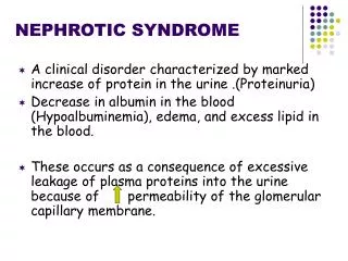
Nephrotic Syndrome (NS). Nephrotic syndrome is a childhood renal disorder occurs as a result of increased glomerular basement membrane permeability, which allows abnormal loss of protein in the urine. It is characterized by: 1. Hyperprotienuria 2. Hypoproteinemia

E N D
Nephrotic Syndrome (NS) Nephrotic syndrome is a childhood renal disorder occurs as a result of increased glomerular basement membrane permeability, which allows abnormal loss of protein in the urine. It is characterized by: • 1. Hyperprotienuria 2. Hypoproteinemia • 3. Hyperlipidemia 4. Generalized edema
Pathophysiology • Increased glomerular permeability results in the passage of larger plasma proteins through the glomerular basement membrane. This results in excess loss of protein (albumin) in the urine (proteinuria) and decreased protein and albumin (hypoalbuminemia) in the bloodstream. • Protein loss in nephrotic syndrome causing hypoalbuminemia results in a change in osmotic pressure, and fluid shifts from the bloodstream into the interstitial tissue (This decrease in blood volume triggers the kidneys to respond by conserving sodium and water, leading to further edema.
Classification: • A-Primary Idiopathic NS (INS): majority The cause is still unclear up to now. Recent 10 years ,increasing evidence has suggested that INS may result from a primary disorder of T– cell function. Accounting for 90% of NS in child. mainly discussed. • B-Secondary NS: NS resulted from systemic diseases, such as anaphylactoid purpura , systemic lupus erythematosus, HBV infection. • C-Congenital NS: rare *1st 3monthe of life ,only treatment renal transplantation
Nephrotic Criteria:- *Massive proteinuria: qualitative proteinuria: 3+ or 4+, quantitative proteinuria : more than 40 mg/m2/hr in children (selective). *Hypo-proteinemia : total plasma proteins < 5.5g/dl and serum albumin : < 2.5g/dl. *Hyperlipidemia: serum cholesterol : > 5.7mmol/L *Edema: pitting edema in different degree
Secondary NS Drug,Toxic,Allegy: mercury, snake venom, vaccine, pellicillamine, Heroin, gold, NSAID, captopril, probenecid, volatile hydrocarbons Infection: APSGN, HBV, HIV, shunt nephropathy, reflux nephropathy, leprosy, syphilis, Schistosomiasis, hydatid disease Autoimmune or collagen-vascular diseases: SLE, Hashimoto’s thyroiditis,, HSP, Vasculitis Metabolic disease: Diabetes mellitus Neoplasma: Hodgkin’s disease, carcinoma ( renal cell, lung, neuroblastoma, breast, and etc) Genetic Disease: Alport syn, Sickle cell disease, Amyloidosis, Congenital nephropathy Others: Chronic transplant rejection, congenital nephrosclerosis
Pathogenesis of Proteinuria:- Increase glomerular permeability for proteins due to loss of negative charged glycoprotein Degree of protineuria:- Mild less than 0.5g/m2/day Moderate 0.5 – 2g/m2/day Sever more than 2g/m2/day Type of proteinuria:- A-Selective proteinuria: where proteins of low molecular weight .such as albumin, are excreted more readily than protein of HMW B-Non selective : LMW(low molecular weight )+HMW(high molecular weight) are lost in urine
pathogenesis of edema:- *Reduction plasma colloid osmotic pressure↓ secondary to hypoalbuminemia Edema and hypovolemia *Intravascular volume↓ antidiuretic hormone (ADH ) and aldosterone(ALD) water and sodium retention Edema *Intravascular volume↓ glomerular filtration rate (GFR)↓ water and sodium retention Edema
Clinical Manifestation:- : 1.Main manifestations: Edema (varying degrees) is the common symptom Local edema: edema in face , around eyes( Periorbital swelling) , in lower extremities. Generalized edema (anasarca), edema in penis and scrotum. 2-Non-specific symptoms: Fatigue and lethargy loss of appetite, nausea and vomiting ,abdominal pain , diarrhea body weight increase, urine output decrease pleural effusion (respiratory distress)
Investigations:- • 1-Urine analysis:- A-Proteinuria: 3-4 + SELECTIVE. b-24 urine collection for protein >40mg/m2/hr for children c- volume: oliguria (during stage of edema formation) d-Microscopically:- microscopic hematuria 20%, large number of hyaline cast
Investigations:- 2-Blood: A-serum protein: decrease>5.5gm/dL , Albumin levels are low (<2.5gm/dL). B-Serum cholesterol and triglycerides:Cholesterol >5.7mmol/L (220mg/dl). C-- ESR↑>100mm/hr during activity phase 3.Serum complemen: Vary with clinical type. 4.Renal function .
General therapy:- • Hospitalization:- for initial work-up and evaluation of treatment. • Activity: usually no restriction , except • massive edema,heavy hypertension and infection. • Diet Hypertension and edema: Low salt diet (<2gNa/ day) only during period of edema or salt-free diet. Severe edema: Restricting fluid intake • Avoiding infection:very important. • Diuresis: Hydrochlorothiazide (HCT) :2mg/kg.d Antisterone : 2~4mg/kg.d Dextran : 10~15ml/kg , after 30~60m, followed by Furosemide (Lasix) at 2mg/kg .
Induction use of albumin:- Albumin + Lasix 1-Severe edema 2-Ascites 3-Pleural effusion 4-Genital edema 5-Low serum albumin
Corticosteroid—prednisone therapy:- Prednisone tablets at a dose of 60 mg/m2/day (maximum daily dose, 80 mg divided into 2-3 doses) for at least 4 consecutive weeks. After complete absence of proteinuria, prednisone dose should be tapered to 40 mg/m2/day given every other day as a single morning dose. The alternate-day dose is then slowly tapered and discontinued over the next 2-3 mo.
Nursing Consideration • Bed rest during the edema with turning regularly to prevent tissue breakdown. • Support edematous areas such as scrotum, abdomen, and legs. • Monitor urine output and the amount of protein in the urine (by dipstick). • Weigh the child daily on the same scale either naked or wearing the same amount of clothing. • Measure pulse rate and blood pressure every 4 hours to detect hypovolemia resulting from excessive fluid shifts. • In cases of severe hypoalbuminemia, intravenous albumin may be administered with lasix
Measures to prevent infection: • Provide good nutrition: • Restrict sodium during edema and steroid therapy. • Give supplementary vitamins and iron as ordered. • Fluid restriction is reserved for children with massive edema. • Encourage protein-rich snacks.
Glomerulonephritis • Glomerulonephritis is an inflammation of the glomerulus. There are two types of glomerulonephritis: Acute poststreptococcal glomerulonephritis (APSGN) occurs as an immune reaction to streptococcal infection of the throat or skin that causing edema, decreases filtering and thus causes urine to be retained. Chronic: An abnormal immune system, bacterial or viral infection, disease or toxin causes progressive dysfunction of the glomerulus over the years
Signs and Symptoms • _ Acute: oliguria, fever, edema of the face and extremities, hypertension and lethargy • _ Chronic: oliguria, hypertension and doesn’t respond to treatment for acute glomerulonephritis • Test Results • Acute: • Hematuria, high specific gravity, proteinuria, elevated KFT • Positive for streptococcal bacteria. • Throat culture: Positive for streptococcal bacteria • Renal ultrasound: Shows enlarged kidneys • Chronic: As the manifestations of acute with two additional symptoms • Show elevated potassium (hyperkalemia) • Ultrasound shows decreased size of kidneys
Treatment • Acute: • Administer antibiotics for 10 days. • Administer diuretics to reduce edema. • Administer corticosteroids to reduce the inflammatory response.
Administer antihypertensive medication to reduce blood pressure. • Low-sodium, low-protein diet and fluid restriction to prevent fluid retention. • Dialysis if the patient experiences renal failure. • Chronic: as the treatment of acute with more focus on • Dialysis or kidney transplant if the patient experiences renal failure. • For emergency hyperkalemia: administer insulin, hypertonic glucose, and calcium gluconate • To remove potassium, administer sodium polystyrene sulfonate (Kayexalate).
Nursing Intervention • Strict intake and output and monitoring daily weights. • Provide a quiet environment. • Monitor vital signs and report changes to the health-care provider. • Explain to the family the importance of a low-salt, low-protein, and fluid restricted diet, and teach the family not to stop administering antibiotics when the child’s condition improves. • Monitor for signs of hyperkalemia (muscle weakness, paresthesia, anorexia, and malaise). • Nursing alert: Monitor for renal failure where urine output <1 mL/kg per hour in infants and <0.5 mL/kg per hour in children, and creatinine, BUN, and urine creatinine clearance are elevated.
Urinary Reflux • Backflow of urine from the bladder into the ureters and kidneys. • Causes: • Infections—20 % of children with UTIs will have reflux with voiding • Anatomical abnormalities • Congenital abnormal insertion of ureters into the bladder. • Treatment: • Antibiotic therapy: may be long term to keep urine sterile (low dose for 6 months) • Surgery to correct problem with insertion of ureters
Nursing care: • Observe and record drainage separately from each catheter. • May have as many as 5 urinary drainage catheters: Each kidney, Ureteral catheters in both ureters and Suprapubic catheter • Encourage fluid intake to maintain hydration • Pain control and antispasmodics to prevent renal colic
Hydrocele • Excessive amounts of fluid in the scrotum-surrounding the testicles. Swollen and painful • Treatment • Often is absorbed with not intervention • Surgery – incision and drainage.