Download
1 / 76
800 likes | 1.5k Views
BURN. Objectives. Describe epidemiology of burn injury Discuss causes of burn Classify burn injury Discuss Pathophysiology of burn Assessment of burn patient Describe treatment plans for burn patient by using ATLS principles Discuss complications of burn. Introduction. Burn

E N D
Objectives • Describe epidemiology of burn injury • Discuss causes of burn • Classify burn injury • Discuss Pathophysiology of burn • Assessment of burn patient • Describe treatment plans for burn patient by using ATLS principles • Discuss complications of burn BURN
Introduction Burn Tissue injury • thermal ( heat, cold) • electrical • Radiation • chemical • coagulative necrosis BURN
Epidemiology • 1% of the world population each year • USA ~ 2.4 million burn injuries/ yr & 10,000 death/yr • UK ~ 250,000 patients treated with burns & 700 deaths/yr. • In Kenya 5,000 deaths/yr • TZ(MNH) 10% of admission in pediatric surgical ward • ??BMC BURN
epid…… Age Scald - < 5 year of age flame, electrical & chemical burn - adult Sex domestic burn - females occupational - males Race No race predilection exists in burn injuries BURN
Risks factors Diseases e.g. epilepsy, diabetes Children< 5years; Elderly > 75 years Cold weather Occupational – electricians/industrial Alcoholism ??Low socioeconomic status BURN
High morbidity and mortality emotional & psychological BURN
Anatomy Skin The epidermis • derived from ectoderm • it can regenerate. The dermis • from mesoderm • cannot re-generate, BURN
AETIOLOGY Thermal injuries Scald Flame Flash Contact Chemical injuries Electrical injuries Radiation injuries Cold injuries BURN
classification • type /cause • body site • degree • size/extent • severity BURN
Class.. - type • Thermal burn • Scald • Flame burn • Contact burn • Flash • Electrical burn • Chemical burn • Radiation burn • Cold burn BURN
Class.. site • Facial burn • Head & neck • Trunk • Limbs • Perineal burn depth • Superficial burn • Epidemal • Dermal • Deep burn • Dermal • Full thickness • Mixed burn BURN
Class.. degree of tissue injury • First degree burn • Second degree burn • 2nd Degree Superficial (superficial Dermal) • 2nd Degree Deep (deep Dermal) • Third degree burn • Fourth degree burn BURN
Class.. Size/Extent Total body surface area (TBSA) burned severity of burn • Minor burn • Moderate burn • Major burn BURN
PATHOPHYSIOLOGY Burn injuries result in:- local response systemic response BURN
Pathophysiology…… LOCAL RESPONSE • Inflammation • Jackson zones (1947) • coagulation /necrosis • Stasis/ischaemia • hyperemia BURN
SYSTEMIC RESPONSE:- Significant burn massive release of inflammatory mediators, both in the wound and other sites. Pathophysiology…… BURN
Follow burn injury , neutrophils ,monocytes & platelets migrate into burn wound • Capillary permeability locally & in distinct organs. • Plasma oncotic pressure • Interstitial oncotic pressure due to increased capillary permeability protein loss edema in burned & un-burned tissues BURN
Biochemical … ↓ tissue perfusion tissue hypoxia anaerobic resp Pyruvate↑ lactic acid metabolic acidosis alter cellular enzymes activity BURN
Biochemical….. ↓ATP↓ Na+Ka+-ATPase ↑↑Na+ intracellular & ↑↑K+ extracellular cellular swelling hyperkalemia ↓ ECF vol. Cell death by necrosis or apoptosis BURN
CVS • ↓Myocardial contractility TNF • ↓ CO due to loss of intravascular vol, ↑ viscocity & ↓cardiac contractility. These changes, coupled with fluid loss from the burn wounds systemic hypotension & end organ hypotension MOD MOF BURN
Respiratory Inflammatory mediators →bronchoconstriction, → ARDS Pulmonary dysfunction • Inhalation injury • Aspiration • Shock • Circumferential thoracic eschar BURN
GIT • mucosal atrophy • changes in the digestive absorption • intestinal permeability Thromboxane A2 prominent mesenteric vasoconstriction ↓gut blood flow compromise gut mucosal intergrity & ↓ immune fxn • Stress (Curling’s) ulcer ( stomach & duodenum). • Acute pseudo-obstruction of the colon (Adynamic ileus) • Acute dilatation of the stomach & colon. • Acalculous cholecystitis BURN
Renal Changes BV &↓ CO RBF GFR ATN ARF BURN
CNS Changes CNS dysfunction in up to 14% of burn patients • Delirium, disorientation Hypoxia • smoke inhalation, • pulmonary edema, • pneumonia BURN
Haematological • Haemoconcentration • Anaemia • Destruction of RBC • Continual loss of RBC for 1 wk • Mild thrombocytopenia (sequestration) early, followed by thrombocytosis (2-4x > normal) by end of the 1st week Persistant thrombocytopenia associated with poor prognosis suspect sepsis • DIC with generalized bleeding can occur shock, sepsis, hypoxia, reperfusion injury BURN
Immunological Innate immunity Skin Cellular Immune Function lymphocyte function Humoral Immune Function IgG & IgA BURN
Metabolic • Ebb phase • Flow phase Catabolic phase Anabolic [recovery phase] BURN
Ebb phase Occurs during the 1st 24 hours • hypothermia • CO & O2 consumption BURN
Catabolic Phase Occurs after 24 hours of burn injury • Mediated through release of catabolic hormones [ eg, catecholamines, glucocorticoids, glucagon ] and other chemical mediators e.g. cytokines, lipid mediators. • ↑ Cardiac output • ↑ Oxygen consumption • ↑ Heat production [hyperthermia] • ↑ BMR • Hyperglycemia • Proteolysis • Peripheral lipolysis BURN
BURNSTRESS Catecolamines CORTISOL GLUCAGON Proteolysis Peripheral Lipolysis Gluconeogenesis AMINO ACIDS GLUCOSE FREE FAT ACIDS BURN
Anabolic / recovery phase Characterized by:- This phase continues for weeks to months after injury Slow re-accumulation of protein and fat BURN
ASSESSMENT OF BURN INJURY Remember • Establish cause. • Associated injuries • During escape from fire. • Explosions throw patient a distance causing internal injuries. • Electrical muscular spasms can cause fractures. • Burns in enclosed space suggest inhalational injury. • Pre-existing disease states, medication, allergies, lung sensitivities. • Establish tetanus immunization status. BURN
Clinical assessment History Physical examination General Local Systemic BURN
history Patient characteristics age , occupation History of injury Time of burn Place of burn Nature of injury Intentional Unintentional Undetermined BURN
History…. • Type of burn • Thermal • Chemical • Electrical • Radiation • Cold • Mechanism of injury • Associated injuries • Associated inhalation injuries • Associated clothing ignition • Whether first aid measures was done at the site of accident BURN
ROS • PMHx ?? Epilepsy, DM, Psychosis • FSHx ??alcohol BURN
General Exam Body weight Shock Level of consciousness Dyspnoea In pain Restless ± gasping Anaemic Dehydration BURN
Physical examination Local examination [assessment of burn wound] • Examine the wound • Body region burned • Extent of burn • Burn depth • Severity of burn Systemic examination • Cardiovascular system • Respiratory system • PA • CNS BURN
Local exam Body region • Head / neck • Upper limbs • Trunk • Lower limbs • Genitalia / Perineal areas BURN
Extent of burn Size of a Burn Injury Total Body Surface Area (TBSA) Burned Palmar Method A quick method to evaluate scattered or localized burns Client’s palm = 1 % TBSA Rule of Nines (Wallace’s) A quick method to evaluate the extent of burns Major body surface areas divided into multiples of nine Modified version for children and infants (Rule of Sevens ) Lund-Browder Method Most Accurate; based on age (growth) Can be used for the adult, children & infants BURN
Burn depth • Superficial (1st Degree) • Partial Thickness Superficial (2nd Degree) Deep ( 2nd Degree) • Full Thickness (3rd Degree) • Deep-Full Thickness (4th degree) BURN
Superficial first degree burn Epidermis Wound Appearance: • Red to pink (light skin) • Mild edema • Dry and no blistering • Pain / hypersensitivity to touch • i.e. Classic sunburn • Desquamation occurs 2-3 days Wound Healing • Wound Healing spontaneous • Duration 3 to 5 days • No scarring / other complications BURN 46
Superficial second degree burn upper 1/3 of dermis • Wound Appearance • Red to pink • Wet and weeping wounds • Thin-walled, fluid-filled blisters • Mild to moderate edema • Extremely painful • Wound Healing • In 2 weeks (spontaneous) • Minimal scarring; minor pigment discoloration may occur BURN
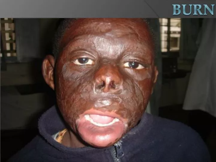
BURN Deep second degree burn deep dermis layer • Wound Appearance • Mottled: Red, pink, to white surface • Moist • Moderate edema • Painful; usually less severe than superficial 2nd Degree superficial. • No blisters • Wound Healing • May heal spontaneously 2-6 weeks • If so Hypertrophic scarring / formation of contractures • Wound Management: • Treatment of choice surgical excision & skin grafting
Full thickness third degree burn entire epidermis and dermisSubcutaneous fat • Wound Appearance • Dry, leathery and rigid • + Eschar (hard and in-elastic) • Red, white, yellow, brown or black • Severe edema • Painless & insensitive to palpation BURN
Wound Healing • No spontaneous healing; • No epidermal or dermal appendages remain, thus must heal by re-epithelialization from the wound edges. • Wound Management: Surgical excision & skin grafting Cx severe scarring/contracture BURN