Download
1 / 17
260 likes | 840 Views
Clonorchiasis Sinensis. Dept. Of Infectious Disease Shengjing Hospital. Intruduction. The disease is a kind of chronic parasitosis caused by clonorchis sinensis , which inhabit human intrahepatic ducts Clinical Manifestation: hepatomegaly, vague pain of upper abdomen, lassitude and tiredness

E N D
Clonorchiasis Sinensis Dept. Of Infectious Disease Shengjing Hospital
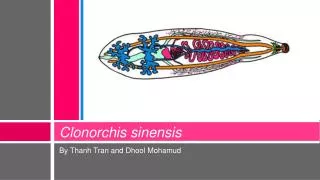
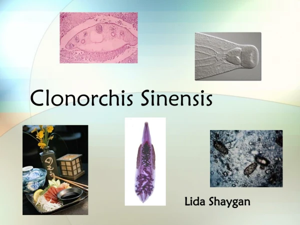
Intruduction • The disease is a kind of chronic parasitosis caused by clonorchis sinensis, which inhabit human intrahepatic ducts • Clinical Manifestation: hepatomegaly, vague pain of upper abdomen, lassitude and tiredness • The infection is acquired by ingestion of raw or inadequately cooked freshwater fish or shrimp
Etiology • Morphology • Adult worm Flat, elongated worm, with the size 10-15×3-5 mm They are monocious, with two suckers The most characteristic feature is branched testis in the posterior third of the body, and relative small ovary before them • Eggs The smallest one of the eggs of human parasites. The sizes are 27.3-35.1×11.7-12.9m Yellow brown operculated eggs,with a fully embryonated miracidia in it
Life Cycle adult worm in human and mammal eaten 1 month eggs into water metacercariae first intermediate host Secocond intermediate host (special snail) (Freshwater fish and shrimp) swallow eggs invade miracidia enter water 2 months cercaria
Epidemiology • Source of infection: patients, infected reservior hosts:cats, dogs, mice, pigs • Route of transmission: the infection is acquired by eating raw or inadequately cooked freshwater fish or shrimp, which are previously infected • Susceptibility: human is generally susceptive, related with the dietary habits
Pathogenesis • Worms mechanical stimulation proliferate inflammatory reaction in the biliary epithelium • The wall of the bile ducts thickened ,fibrous tissue around the bile duct, and worm obstruction cause cholestasis • When bacteria infection occur, cause cholecystitis, cholangeitis, sometimes cholelithiasis happens
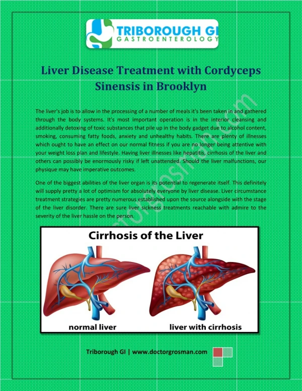
Pathology • Intrahepatic bile ducts expansion, hepatomegaly, necrosis of liver tissue • Continuous severe infection may cause liver cirrhosis • Persistent Cholestasis cause biliary liver cirrhosis • Associate with cholangiocarcinoma and hepatic carcinoma
Clinical Manifestation • Incubation period: 1-2 months • Most person with mild clonorchis sinensis infections are asymptomatic, only eggs can be found in the feces • Severe infections: onset is insidious, with intestinal manifestations like viral hepatitis, hepatomegaly, neurasthenia, person with heavy worm loads may suffer from biliary angina and obstructive icteric
Clinical Manifestation • Acute symptoms appear when the primary infection is heavy: sudden onset, chill, high fever, slight jaundice, hepatomegaly, eosinophilia, a few patients have splenomegaly, and weeks later, enter chronic stage • Continuous reinfection: cirrhosis and portal hypertension. In children may cause malnutrition growth development disturbance, even dwarf
Complications • Acute or chronic cholecystitis, cholangeitis and cholelithiasis are the most common complications • Portal liver cirrhosis: portal hepertension result in upper gastrointestinal bleeding • Cholestatic cirrhosis • Pancreatitis • Primary carcinoma of the liver and cholangiocarcinoma
Laboratory Findings • Blood routine test: eosinophilia, anemia in severe infection • Eggs examination: simple smear feces to find eggs Stool concentration technique may increase the positive rate Duodenal aspiration: raise the chance of finding eggs
Laboratory Findings • Immunological Test • Skin test: positive rate 97.9%, 99.5% coincide with the result of the feces • PHA: positive rate 53.7%, 80% coincide with the result of the feces • ELISA: positive rate 98.3%, 93.5% coincide with the result of the feces
Diagnosis • Epidemiologic date: living in or come from the endemic area The history of eating raw or inadequately cooked freshwater fish and shrimp • Clinical date: gastrointestinal symptoms, hepatomegaly, neurasthenia, cholangoitis, cholecystitis, etc. • Laboratory findings: Discovery ofcharacteristic eggs in feces or by duodenal aspiration come to accurate diagnosis Eosinophilia and positive immunologic test support the diagnosis
Differential Diagnosis • Viral hepatitis • Liver cirrhosis of other origins • Primary carcinoma of the liver • Fasciolopsiasis • Other specie of flukes infection
Prognosis • Good of the mild infection • Co-infection with viral hepatitis may make the disease severe
Treatment • Pathogenic Treatment • Praziquantel is the best choice of drug for the therapy • Dose: 15-25mg/kg, three times a day, for 2 days, the total dose is 90-150mg/kg • Another choice of drug is Albendazole • Heteropathy Treatment
Prevention • Control of the source of infection: Treat the patients and domestic animal(cats and dogs, etc.) at the same time. • Cut off the route of transmission: Avoid of eating inadequately cooked freshwater fish and shrimp Sanitary disposal of the excreta Avoid of drinking raw water