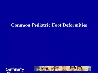
Common Pediatric Foot Deformities
1.45k likes | 1.66k Views
Dive into the evaluation and causes of common foot and leg deformities in children, including bow legs, knock knees, and rotational issues, with detailed clinical examinations and diagnostic approaches. Learn when to refer for surgery and how to address angular and rotational deformities.

Common Pediatric Foot Deformities
E N D
Presentation Transcript
Common Orthopedic Problemsin Children • Angular deformities of LL: • Bow legs. • Knock knees. • Rotational deformities of LL: • In-toeing. • Ex-toeing. • Leg aches. • CDH. • Feet problems. • Irritable hip.
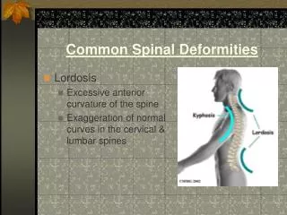
Bow legs Knock knees Angular DeformitiesNomenclature Genu Varus Genu Valgus
Angular DeformitiesRange of Normal Varies With Age • During first year : Lateral bowing of Tibiae • During second year : Bow legs (knees & tibiae) • Between 3 – 4 years : Knock knees
Angular DeformitiesEvaluation Should differentiate between “physiologic” and “pathologic” deformities
Physiologic Pathologic Angular DeformitiesEvaluation • Symmetrical • Asymmetrical • Mild – moderate • Severe • Progressive • Regressive • Generalized • Localized • Expected for age • Not expected for age
Physiologic Pathologic Angular DeformitiesCauses • Normal – for age • Rickets • Exaggerated : • Endocrine disturbance • Metabolic disease - Overweight • Injury to Epiphys. Plate • Infection / Trauma - Early wt. bearing - Use of walker? • Idiopathic
Angular DeformitiesEvaluation Symmetrical deformity
Angular DeformitiesEvaluation Asymmetrical Deformity
Angular DeformitiesEvaluation Generalized deformity
Angular DeformitiesEvaluation Localized deformity Blount’s
Angular DeformitiesEvaluation Localized deformity Rickets
Angular DeformitiesEvaluation Measure Angulation ( standing / supine ) in bow legs / genu varum Inter-condylar distance
Angular DeformitiesEvaluation Measure Angulation ( standing / supine ) in knock knees /genu valgum Inter- malleolar distance
Angular DeformitiesEvaluation Measure Angulation Use goneometer measures angles directly
Angular DeformitiesEvaluation • Serum Calcium / Phosphorous ? • Serum Alkaline Phosphatase • Serum Creatinine / Urea – Renal function Investigations / Laboratory
Angular DeformitiesEvaluation X-ray when severe or possibly pathologic • Standing AP film • long film ( hips to ankles )with patellae directed forwards • Look for diseases : • Rickets / Tibia vara (Blount’s) / Epiphyseal injury.. • Measure angles. Investigations / Radiological
Angular DeformitiesEvaluation Investigations / Radiological Medial Physeal Slope Femoral-Tibial Axis
Angular DeformitiesWhen To Refer ? • Pathologic deformities: Asymmetrical. Localized. Progressive. Not expected for age. • Exaggerated physiologic deformities: Definition ?
Rotational LL Deformities • Frequently seen. • Concerns parents. • Frequently prompts varieties of treatment. ( often un-necessary / incorrect ) In-toeing / Ex-toeing
Rotational Deformities • Level of affection : Femur Tibia Foot
Rotational DeformitiesFemur Ante-version = more medial rotation Retro-version = more lateral rotation
Rotational DeformitiesNormal Development • Femur : Ante-version : • 30 degrees at birth. • 10 degrees at maturity. • Tibia : Lateral rotation : • 5 degrees at birth. • 15 degrees at maturity.
Rotational DeformitiesNormal Development Both Femur and Tibia laterally rotate with growth in children • Medial Tibial torsion and Femoral ante-version improve ( reduce ) with time. • Lateral Tibial torsion usually worsens with growth.
Rotational DeformitiesClinical Examination Rotational Profile • At which level is the rotational deformity? • How severe is the rotational deformity? • Four components: 1- Foot propagation angle. 2- Assess femoral rotational arc. 3- Assess tibial rotational arc. 4- Foot assessment.
Rotational DeformitiesClinical Examination Rotational Profile 1- Foot propagation angle – Walking Normal Range: +10o _10o ? In Eastern Societies +25o _10o
Rotational DeformitiesClinical ExaminationRotational Profile2- Assess Femoral Rotational Arc Supine Extended
Rotational DeformitiesClinical ExaminationRotational Profile2- Assess Femoral Rotational Arc Supine flexed
Rotational DeformitiesClinical Examination Rotational Profile 3- Tibial Rotational Arc Thigh-foot angle in prone foot position is critical leave to fall into natural position
Rotational DeformitiesClinical ExaminationRotational Profile4- Foot assessment • Metatarsus adductus • Searching big toe • Everted foot • Flat foot
Rotational DeformitiesCommon Presentations Infants • Out-toeing : Normal • seen when infant positioned upright ( usually hips laterally rotate in-utero ) • Metatarsus adductus : • medial deviation of forefoot • 90 % resolve spontaneously • casting if rigid or persists late in 1st year
Rotational DeformitiesCommon Presentations Toddlers • In-toeing most common during second year. ( at beginning of walking ) • Causes : • medial tibial torsion. • metatarsus adductus. • abducted great toe.
Rotational DeformitiesCommon Presentations Toddlers - Medial Tibial Torsion • The commonest cause of in-toeing • Observational management is best • Avoid special shoes / splints / braces • unnecessary, ineffective, interferes with activity and cause psychological and behavioral problems.
Rotational DeformitiesCommon Presentations • Serial casting is effective in this age-group • Usually correctable by casting up to 4 years Toddler - Metatarsus Adductus
Rotational DeformitiesCommon Presentations • Dynamic deformity • Over-pull of Abductor Hallucis Muscle during stance phase Toddlers - Abducted Great Toe • Spontaneously resolve - no treatment
Rotational DeformitiesCommon PresentationsChild • In-toeing : due to medial femoral torsion • Out-toeing : in late childhood lateral femoral / tibial torsion
Rotational DeformitiesCommon PresentationsChildMedial Femoral Torsion • Usually: - starts at 3 - 5 years, - peaks at 4 – 6 years, - then resolves spontaneously. • Girls > boys. • Look at relatives - family history – normal. • Treatment usually not recommended. • If persists > 8 years and severe, may need surgery.
Rotational DeformitiesCommon Presentation Medial Femoral Torsion (Ante-version) • Stands with knees medially rotated (kissing patellae). • Sits in W position. • Runs awkwardly (egg-beater). Family History
Rotational DeformitiesCommon PresentationsChildLateral Tibial Torsion • Usually worsens. • May be associated with knee pain (patellar) specially if LTT is associated with MFT. ( knee medially rotated and ankle laterally rotated )
Rotational DeformitiesCommon PresentationsChildMedial Tibial Torsion • Less common than LTT in older child • May need surgery if : • persists > 8 year, • and causes functional disability
Rotational DeformitiesManagement • Challenge : dealing effectively with family • In-toeing : spontaneously corrects in vast majority of children as LL externally rotates with growth - Best Wait !
Rotational DeformitiesManagement Convince family that only observation is appropriate • < 1 % of femoral & tibial torsional deformities fail to resolve and may require surgery in late childhood.
Rotational DeformitiesManagement • Attempts to control child’s walking, sitting and sleeping positions is impossible and ineffective cause frustration and conflicts. • She wedges and inserts : ineffective. • Bracing with twisters :ineffective - and limits activity. • Night splints : better tolerated - ? Benefit.
Rotational DeformitiesManagement Shoe wedges Ineffective Twister cables Ineffective
Rotational DeformitiesWhen To Refer ? • Severe & persistent deformity. • Age > 8-10y. • Causing a functional dysability. • Progressive.
Rotational DeformitiesManagementWhen Is Surgery Indicated ? • In older child ( > 8 – 10 years ). • Significantfunctional disability. • Notprophylactic !
Leg Aches / Growing Pains • Incidence : 15-30 % of children. • More In girls / At night / In LL. • Diagnosis is made by exclusion.